


Key Takeaways
Patellar luxation is a common orthopedic condition where the kneecap dislocates from its normal position in the femoral groove
- Patellar luxation is a common orthopedic condition where the kneecap dislocates from its normal position in the femoral groove
- Small breed dogs like Yorkshire Terriers, Toy Poodles, and Chihuahuas are most commonly affected by medial patellar luxation
- The condition is graded from I to IV based on severity, with higher grades requiring surgical intervention
- Early diagnosis and treatment prevent progression to arthritis and other knee complications
- Surgical correction offers excellent outcomes when performed before severe joint damage occurs
Small breed dogs like Yorkshire Terriers, Toy Poodles, and Chihuahuas are most commonly affected by medial patellar luxation
The condition is graded from I to IV based on severity, with higher grades requiring surgical intervention
Early diagnosis and treatment prevent progression to arthritis and other knee complications
Surgical correction offers excellent outcomes when performed before severe joint damage occurs
When your dog suddenly lifts a hind leg mid-stride, shakes it, and continues walking normally, you may be witnessing the characteristic sign of patellar luxation. This orthopedic condition affects approximately 7% of puppies and represents one of the most common knee joint disorders in dogs. Understanding patellar luxation—from its underlying causes to modern surgical treatments—empowers dog owners to recognize early signs and seek appropriate veterinary care before permanent damage occurs.
What is Patellar Luxation?
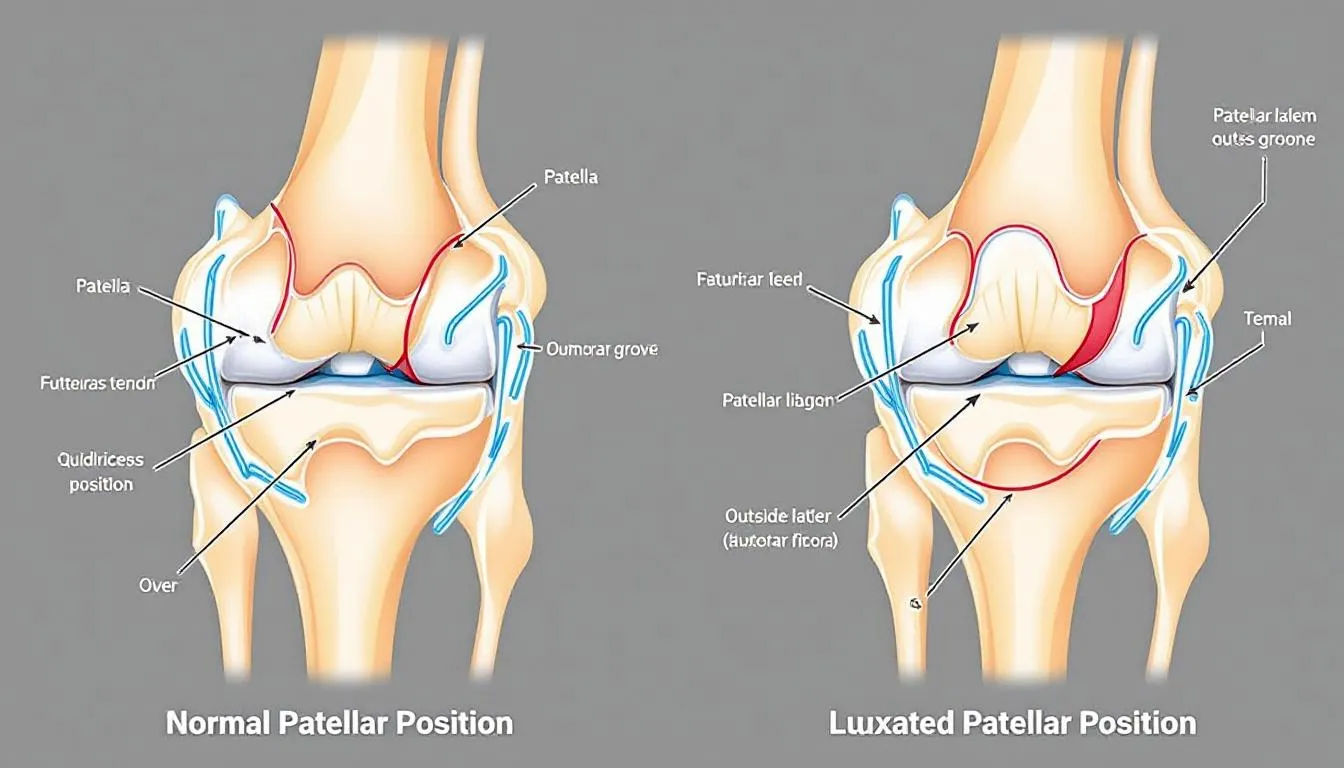
Patellar luxation occurs when the patella (kneecap) dislocates from its normal position within the femoral groove of the thigh bone. In healthy dogs, the patella functions as a crucial component of the extensor mechanism, acting as a fulcrum that allows the quadriceps muscle to efficiently extend the knee joint during movement.
The patella is a small sesamoid bone embedded within the patellar tendon, which connects the quadriceps muscle group to the tibial tuberosity on the shin bone. During normal knee function, the patella glides smoothly within the trochlear groove of the distal femur, maintaining proper alignment of the quadriceps mechanism.
Luxating patella can occur in two primary directions: medial patellar luxation (toward the inside of the leg) and lateral patellar luxation (toward the outside). Medial luxation is far more common, particularly in small breed dogs, while lateral luxation occurs more frequently in large breed dogs and may be associated with hip dysplasia or trauma.
The stifle joint’s stability depends on the coordinated function of bones, soft tissues, and the patellar ligament. When the patella luxates, it disrupts this delicate balance, leading to abnormal stress on surrounding structures and potential long-term complications including damage to the cranial cruciate ligament.
Causes and Risk Factors
The cause of patellar luxation varies significantly between small and large breed dogs, with genetic predisposition playing the primary role in miniature breed dogs. Congenital anatomical abnormalities affect the normal development of the quadriceps mechanism, creating an environment where the patella cannot maintain its proper position within the femoral groove.
In small breed dogs, several skeletal abnormalities contribute to patellar luxation development:
- Shallow or absent medial trochlear ridge
- Abnormally shaped femurs with internal rotation
- Valgus deformity of the lower limb
- Malposition of the tibial crest
- Patella alta (high-riding kneecap)
Shallow or absent medial trochlear ridge
Abnormally shaped femurs with internal rotation
Valgus deformity of the lower limb
Malposition of the tibial crest
Patella alta (high-riding kneecap)
Yorkshire Terriers, along with other toy breeds, demonstrate a strong genetic component to this condition, with certain bloodlines showing significantly higher incidence rates. The inheritance pattern suggests multiple genes influence the development of anatomical abnormalities that predispose to luxating patella.
Large breed dogs typically experience lateral luxation occurs due to different anatomical factors. Hip dysplasia frequently accompanies lateral patellar luxation in these dogs, creating a cascade of compensatory changes throughout the hind limbs. Femoral varus (inward bowing) and external rotation of the femoral neck contribute to lateral displacement of the quadriceps muscle relative to its normal alignment.
Traumatic luxation can affect dogs of any size following significant injury to the stifle joint. Vehicle accidents, falls, or direct trauma may tear soft tissue restraints, allowing the patella to luxate even in dogs with normal anatomical structure.
Grading System and Severity
Veterinarians classify patellar luxation using a standardized grading system that helps determine appropriate treatment and prognosis. The grades of patellar luxation range from I to IV, with each grade representing increasing severity and functional impairment.
Grade I Patellar Luxation The patella remains in its normal position during regular activity but can be manually luxated during physical examination. Most dogs with grade I patellar luxation show no significant clinical signs and may not require immediate intervention. The patella returns automatically to the femoral groove when manual pressure is released.
Grade II Patellar Luxation The patella luxates spontaneously during knee flexion or with minimal manual pressure. Dogs with grade ii patellar luxation often demonstrate the characteristic “skipping” lameness, briefly lifting the affected limb before returning to normal gait. The patella can be manually reduced to its normal position and typically stays there during extension.
Grade III Patellar Luxation The patella remains luxated most of the time but can be manually repositioned into the femoral groove. However, it immediately re-luxates when manual pressure is removed or when the dog flexes the knee joint. Dogs affected with grade III luxation show persistent lameness and may develop a bow-legged stance.
Grade IV Patellar Luxation The patella is permanently luxated and cannot be manually repositioned into the trochlear groove. This represents severe patellar luxation with significant deformity of the entire limb. Dogs experience permanent lameness and substantial functional impairment.
Bilateral patellar luxations occur in approximately 50% of affected dogs, though the severity may differ between legs. During routine physical examination, veterinarians assess both knees to determine if one or both knees are affected and grade each limb independently.
Clinical Signs and Symptoms
The clinical presentation of patellar luxation varies dramatically based on the grade of severity and whether the condition affects one or both knees. Early recognition of symptoms allows for timely intervention before the development of secondary complications.
Dogs with intermittent patellar luxation typically exhibit a characteristic “skipping” gait where the kneecap slides out of position, causing sudden three-legged lameness. The dog lifts the affected limb for several steps, often shaking or extending the leg to help the patella return to its normal position. Once repositioned, the dog resumes normal weight-bearing until the next luxation episode.
As the condition progresses, episodes of lameness become more frequent and may last longer. Dogs with persistent lameness often develop compensatory movement patterns that place additional stress on other joints. The affected knee may appear swollen, and owners might notice the dog reluctant to jump, climb stairs, or engage in vigorous play.
In severe cases where the patella luxates permanently, dogs develop characteristic postural changes. Medial luxation typically causes a bow-legged appearance with internal rotation of the affected limb. The dog may walk with a crouched gait, keeping the knee partially flexed to minimize discomfort.
Significant clinical signs warranting immediate veterinary attention include:
- Persistent lameness lasting more than 24 hours
- Visible swelling or heat around the knee joint
- Complete inability to bear weight on the affected limb
- Signs of pain when touching or manipulating the knee
- Progressive worsening of gait abnormalities
Persistent lameness lasting more than 24 hours
Visible swelling or heat around the knee joint
Complete inability to bear weight on the affected limb
Signs of pain when touching or manipulating the knee
Progressive worsening of gait abnormalities
Pain levels vary considerably between individual dogs. Some animals with severe anatomical deformities show remarkable tolerance, while others with mild luxation experience substantial discomfort. Most dogs adapt their activity level to minimize symptoms, which can mask the progression of joint damage.
Diagnosis and Examination
Accurate diagnosis of patellar luxation requires a systematic approach combining physical examination techniques with appropriate imaging studies. The condition can usually be patellar luxation diagnosed through careful palpation and manipulation of the stifle joint, though additional testing helps assess the extent of anatomical abnormalities.
During physical examination, the veterinarian positions the dog in lateral recumbency to properly assess patella mobility. With the knee joint in extension, gentle pressure applied to the patella determines whether it can be displaced from the femoral groove. The examiner notes the direction of luxation (medial or lateral), the ease of displacement, and whether the patella returns spontaneously to its normal position.
Palpation of the affected knee reveals important information about joint stability and associated changes. Thickening of the joint capsule, muscle atrophy of the quadriceps muscle, and crepitus (grinding sensation) may indicate chronic changes and secondary arthritis development.
Radiographic evaluation provides essential information about bone structure and joint alignment. Standard orthogonal views of the stifle joint help identify:
- Depth and shape of the trochlear groove
- Position of the tibial tuberosity
- Presence of femoral varus or other angular limb deformities
- Signs of degenerative joint disease
- Concurrent orthopedic conditions
Depth and shape of the trochlear groove
Position of the tibial tuberosity
Presence of femoral varus or other angular limb deformities
Signs of degenerative joint disease
Concurrent orthopedic conditions
Advanced imaging including CT or MRI may be recommended for complex cases requiring surgical planning. Three-dimensional reconstruction helps surgeons visualize the exact anatomical abnormalities and plan corrective procedures with greater precision.
The veterinarian also evaluates both hind limbs for bilateral involvement and screens for related conditions. Hip dysplasia commonly accompanies lateral luxation in large breed dogs, while cranial cruciate ligament injury may develop secondary to chronic patella instability.
Conservative Treatment Options
Conservative management of patellar luxation focuses on minimizing symptoms and preventing progression of joint damage. This approach is most appropriate for dogs with grade I patellar luxation showing minimal clinical signs or for owners who cannot pursue surgical options.
Weight management represents the cornerstone of conservative treatment. Excess body weight increases stress on the knee joint and exacerbates luxation episodes. Dogs should maintain a lean body condition where ribs are easily palpable and a visible waist is apparent when viewed from above. Even a 10% reduction in body weight can significantly improve comfort and function.
Anti-inflammatory medications, particularly NSAIDs, help manage pain and reduce joint inflammation. These medications should be used under veterinary supervision with appropriate monitoring for potential side effects. The goal is to provide comfort while allowing the dog to maintain reasonable activity levels.
Physical rehabilitation and controlled exercise modification can strengthen the quadriceps muscle and improve joint stability. Swimming and underwater treadmill therapy provide excellent low-impact exercise that builds muscle strength without stressing the knee joint. Specific exercises targeting hip and core stability may help compensate for altered biomechanics.
Joint supplements containing glucosamine, chondroitin sulfate, and omega-3 fatty acids may support cartilage health and reduce inflammation. While these supplements cannot correct the underlying anatomical problem, they may slow the progression of arthritis in dogs with chronic luxation.
Activity restriction helps prevent acute luxation episodes and reduces cumulative joint damage. Dogs should avoid high-impact activities like jumping, agility training, and intense play sessions. Leash walks on flat surfaces provide safe exercise while maintaining muscle tone.
Conservative treatment success depends on early intervention and owner compliance. Regular veterinary monitoring allows for adjustment of treatment protocols and early identification of cases requiring surgical intervention.
Surgical Treatment Approaches
Surgical correction of patellar luxation aims to restore normal alignment of the quadriceps mechanism and provide stable patella tracking within the femoral groove. The specific surgical techniques chosen depend on the grade of luxation, anatomical abnormalities present, and the dog’s size and activity level.
Patellar luxation surgical treatments have evolved significantly, with modern approaches offering excellent success rates when performed before severe joint damage occurs. The timing of surgical intervention is crucial—early correction prevents the development of arthritis and maintains normal joint function throughout the dog’s life.
Pre-surgical planning involves detailed assessment of all anatomical abnormalities contributing to the luxation. Advanced imaging helps surgeons identify the specific combination of procedures needed to correct patellar luxation and restore normal alignment. Most cases require multiple surgical techniques performed simultaneously for optimal results.
The primary goals of surgical treatment include:
- Realigning the quadriceps mechanism to center the patella
- Deepening the trochlear groove to improve patella stability
- Correcting angular limb deformities that contribute to luxation
- Balancing soft tissue tensions around the knee joint
- Preventing recurrence while maintaining normal joint function
Realigning the quadriceps mechanism to center the patella
Deepening the trochlear groove to improve patella stability
Correcting angular limb deformities that contribute to luxation
Balancing soft tissue tensions around the knee joint
Preventing recurrence while maintaining normal joint function
Tibial Tuberosity Transposition
Tibial tuberosity transposition represents the most commonly performed procedure to correct patellar luxation. This surgical technique involves cutting the tibial crest (where the patellar tendon inserts) and repositioning it to properly align the extensor mechanism.
During the procedure, the surgeon carefully cuts through the bone at the tibial tuberosity, preserving the attachment of the patellar tendon. The bone fragment is then moved medially (for lateral luxation) or laterally (for medial luxation) to center the pull of the quadriceps muscle group over the femoral groove.
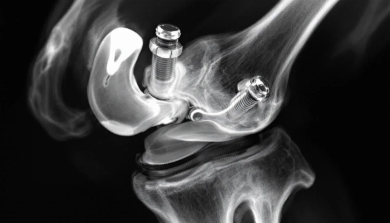
The repositioned tuberosity is stabilized using small pins or screws, allowing the bones heal over a 6-8 week period. This technique offers several advantages over soft tissue procedures, as bone healing provides permanent correction that doesn’t stretch or fail over time.
Post-operative care requires strict exercise restriction during the healing phase. Dogs must be confined to prevent running, jumping, or other activities that could disrupt the healing bone. Most patients show significant improvement in gait within 2-3 weeks, though complete healing takes 6-8 weeks.
Femoral Osteotomy Procedures
Severe cases of patellar luxation may require correction of angular limb deformities through femoral osteotomy. These procedures involve cutting the thigh bone and repositioning it to correct valgus deformity, femoral varus, or excessive internal rotation that contributes to luxation.
Wedge osteotomy techniques remove a precisely calculated piece of bone to achieve the desired angular correction. The bone segments are then stabilized using plates and screws, which remain in place permanently. Advanced planning using CT imaging ensures accurate correction of complex three-dimensional deformities.
Dome osteotomy offers an alternative approach for multi-directional corrections. This technique allows simultaneous correction of angular and rotational deformities through a single curved bone cut. Computer-assisted planning helps achieve precise corrections while preserving limb length.
Recovery from femoral osteotomy requires 8-12 weeks of restricted activity while the bone heals. These procedures are typically reserved for large breed dogs with severe deformities or cases where simpler techniques have failed.
Trochlear Groove Deepening
Dogs with shallow or absent trochlear grooves benefit from surgical deepening to improve patella stability. Several techniques are available, with the choice depending on the severity of groove abnormalities and the presence of articular cartilage.
Recession sulcoplasty involves carefully removing bone from the bottom of the trochlear groove while preserving the overlying articular cartilage. This technique deepens the groove without damaging the smooth joint surface, making it ideal for younger dogs with mild to moderate groove abnormalities.
Wedge or block trochleoplasty techniques involve removing a section of bone from the groove and replacing it at a deeper level. These procedures can achieve more dramatic deepening but may require removal of some articular cartilage, potentially increasing the risk of future arthritis.
The surgical approach must balance the need for adequate groove depth with preservation of normal joint anatomy. Over-deepening the groove can create new problems, making precise technique essential for optimal outcomes.
Soft Tissue Reconstruction
Soft tissue procedures complement bony corrections by balancing the tensions around the knee joint. These techniques involve releasing tight structures on one side while tightening loose tissues on the opposite side to center the patella within the groove.
For medial luxation, surgeons typically release the tight medial joint capsule and fascial structures that pull the patella inward. The lateral joint capsule and surrounding soft tissue are then tightened to provide lateral restraint and prevent re-luxation.
Lateral luxation requires the opposite approach, with release of lateral structures and tightening of medial soft tissue. The goal is to restore normal patella tracking without over-constraining joint motion.
Soft tissue reconstruction alone is rarely sufficient for significant luxation but provides important support when combined with bony procedures. The techniques must be precisely balanced to avoid creating excessive tension that could limit joint motion or cause patella to luxate in the opposite direction.
Post-Operative Care and Recovery
Successful recovery from patellar luxation surgery requires careful attention to post-operative restrictions and rehabilitation protocols. The initial healing phase is critical for achieving optimal long-term outcomes and preventing complications.
During the first 6-8 weeks following surgery, strict exercise restriction is mandatory. Dogs must be confined to prevent running, jumping, or other high-impact activities that could disrupt healing tissues or implants. Crate rest or confinement to a small room helps ensure compliance with activity restrictions.
Short leash walks for elimination purposes are permitted, but the dog should not be allowed to play, interact with other pets, or engage in any vigorous activity. Stairs should be avoided, and the dog should be carried when necessary to prevent jumping on or off furniture.
Pain management during the recovery period typically involves a combination of medications including NSAIDs and sometimes additional analgesics for the first few days after surgery. Ice packs applied for 10-15 minutes several times daily can help reduce swelling and provide comfort.
Physical rehabilitation should begin early under professional guidance. Initial therapy focuses on maintaining joint range of motion through gentle passive exercises. As healing progresses, controlled weight-bearing exercises and strengthening activities help restore normal function.
Swimming and underwater treadmill therapy provide excellent low-impact exercise once initial healing is complete. These activities help rebuild muscle strength while protecting the surgical site from excessive stress.
Regular recheck appointments allow the veterinarian to monitor healing progress and adjust restrictions as needed. Radiographs may be taken to confirm proper implant position and bone healing before clearing the dog for normal activity.
Most dogs show significant improvement in gait within 2-3 weeks of surgery, though complete healing and return to full activity typically takes 3-4 months. Patience during the recovery period is essential for achieving the best possible outcome.
Prognosis and Long-term Outcomes
The prognosis for dogs with patellar luxation is generally excellent when appropriate treatment is instituted before severe joint damage occurs. Early surgical intervention offers the best chance for return to normal function and prevention of long-term complications.
Success rates for surgical correction vary by grade and complexity but generally exceed 90% for properly selected cases. Dogs with grade I and II luxation typically achieve complete resolution of clinical signs and return to full activity. More severe cases may show significant improvement but might retain some degree of gait abnormality.
Several factors influence long-term outcomes:
- Grade of luxation : Higher grades carry increased risk of complications and reduced success rates
- Age at surgery : Younger dogs typically heal faster and achieve better results
- Concurrent conditions : Hip dysplasia or cruciate ligament injury may affect overall prognosis
- Owner compliance : Adherence to post-operative restrictions significantly impacts success
Grade of luxation : Higher grades carry increased risk of complications and reduced success rates
Age at surgery : Younger dogs typically heal faster and achieve better results
Concurrent conditions : Hip dysplasia or cruciate ligament injury may affect overall prognosis
Owner compliance : Adherence to post-operative restrictions significantly impacts success
Potential complications include re-luxation, implant failure, and infection, though these occur in less than 10% of cases when surgery is performed by experienced surgeons. Arthritis development may still occur in cases where significant cartilage damage existed before surgery.
Most dogs affected by patellar luxation can return to normal activity levels including agility, hiking, and other athletic pursuits following successful surgical correction. The key is early recognition and treatment before irreversible joint damage occurs.
Dogs that undergo timely surgical treatment typically enjoy normal lifespans with excellent quality of life. Regular veterinary monitoring helps identify any potential issues early and maintain optimal joint health throughout the dog’s life.
Prevention and Genetic Considerations
Prevention of patellar luxation relies primarily on responsible breeding practices, as the condition has a strong genetic component in most affected breeds. Breeders play a crucial role in reducing the incidence of this condition through careful selection and genetic counseling.
Breeding recommendations include:
- Screening all breeding animals for patellar luxation before reproduction
- Avoiding breeding dogs with grade II or higher luxation
- Maintaining detailed records of offspring to track inheritance patterns
- Working with veterinary geneticists to understand breed-specific risks
Screening all breeding animals for patellar luxation before reproduction
Avoiding breeding dogs with grade II or higher luxation
Maintaining detailed records of offspring to track inheritance patterns
Working with veterinary geneticists to understand breed-specific risks
The Orthopedic Foundation for Animals (OFA) provides patellar luxation screening and certification programs that help breeders make informed decisions. Dogs receiving OFA certification have been examined by qualified veterinarians and found to have normal patella function.
Environmental factors may also influence the development or severity of patellar luxation. Maintaining appropriate body weight during growth helps reduce stress on developing joints. Avoiding excessive exercise or high-impact activities in young, growing dogs may help prevent traumatic luxation in susceptible individuals.
Early screening of puppies in high-risk breeds allows for early detection and intervention. Veterinarians can assess puppies as young as 8 weeks of age, though definitive grading may require re-examination as the dog matures.
Owner education about recognizing early signs of patellar luxation enables prompt veterinary consultation and appropriate treatment. Understanding breed-specific risks helps owners make informed decisions about activity levels and preventive care.
FAQ
Can patellar luxation heal on its own without surgery?
Grade I luxations may not require surgery if the dog shows no clinical signs, but most cases need intervention to prevent progression and arthritis development. Conservative management can help manage symptoms, but it cannot correct the underlying anatomical abnormalities that cause the condition. Without treatment, patellar luxation typically worsens over time, leading to permanent joint damage and chronic pain.
How long does recovery take after patellar luxation surgery?
Initial healing takes 6-8 weeks with exercise restriction, followed by gradual return to normal activity over 3-4 months with proper rehabilitation. The exact timeline depends on the surgical procedures performed, the dog’s age and overall health, and compliance with post-operative restrictions. Most dogs show significant improvement in their gait within 2-3 weeks of surgery, though complete healing requires patience and careful monitoring.
Will my dog be able to participate in agility or high-impact activities after surgery?
Most dogs can return to full activity including agility after successful surgery and complete recovery, though this depends on the grade of luxation and any concurrent conditions. Dogs with grade I or II luxation that receive timely surgical correction typically achieve complete functional recovery. However, dogs with severe pre-existing arthritis or other joint problems may have some activity limitations even after successful surgery.
Is patellar luxation painful for dogs?
Yes, especially in higher grades where the patella is frequently or permanently displaced, causing cartilage wear and eventually arthritis if left untreated. However, pain levels vary significantly between individual dogs. Some animals show remarkable tolerance even with severe deformities, while others experience substantial discomfort with mild luxation. The intermittent nature of grade I and II luxation means pain often comes and goes with luxation episodes.
Can patellar luxation affect both legs simultaneously?
Approximately 50% of dogs with patellar luxation have bilateral involvement, though the severity may differ between legs. When both knees are affected, they may not necessarily be the same grade or require the same treatment approach. Some dogs may have grade II luxation in one leg and grade I in the other, requiring individualized treatment plans for each limb. Bilateral involvement is particularly common in breeds with strong genetic predisposition to the condition.


