


Key Takeaways
- Canine pyoderma is a bacterial skin infection primarily caused by Staphylococcus pseudintermedius, affecting over 90% of cases and requiring prompt veterinary diagnosis
- The condition is classified into three types by depth: surface pyoderma (outermost layer), superficial pyoderma (epidermis and hair follicles), and deep pyoderma (dermis and subcutaneous tissue)
- Topical antimicrobial therapy is the preferred first-line treatment for surface and superficial pyoderma, while systemic antibiotics are reserved for deep infections or treatment failures
- Pyoderma is always secondary to underlying conditions like allergies, parasites, or endocrine disorders, making identification and management of root causes essential for preventing recurrence
- Antibiotic resistance, particularly methicillin-resistant Staphylococcus pseudintermedius (MRSP), is a growing concern requiring culture-guided therapy and antimicrobial stewardship
Canine pyoderma is a bacterial skin infection primarily caused by Staphylococcus pseudintermedius, affecting over 90% of cases and requiring prompt veterinary diagnosis
The condition is classified into three types by depth: surface pyoderma (outermost layer), superficial pyoderma (epidermis and hair follicles), and deep pyoderma (dermis and subcutaneous tissue)
Topical antimicrobial therapy is the preferred first-line treatment for surface and superficial pyoderma, while systemic antibiotics are reserved for deep infections or treatment failures
Pyoderma is always secondary to underlying conditions like allergies, parasites, or endocrine disorders, making identification and management of root causes essential for preventing recurrence
Antibiotic resistance, particularly methicillin-resistant Staphylococcus pseudintermedius (MRSP), is a growing concern requiring culture-guided therapy and antimicrobial stewardship
Bacterial skin infections represent one of the most common dermatological challenges in veterinary medicine, with pyoderma in dogs affecting an estimated 15-20% of all canine dermatological presentations. This comprehensive guide explores the complexities of canine pyoderma, from its underlying mechanisms to evidence-based treatment protocols that prioritize both clinical efficacy and antimicrobial stewardship.
Understanding pyoderma requires recognizing its fundamentally secondary nature—this bacterial skin infection never occurs in isolation but always develops as a consequence of predisposing factors that compromise the skin barrier or immune system. This principle guides both diagnostic approaches and long-term management strategies essential for preventing recurrent infections.
What Is Canine Pyoderma?
Canine pyoderma represents a bacterial skin infection characterized by the presence of pus within skin tissues, with the term deriving from the Greek words “pyo” (pus) and “derma” (skin). Unlike human skin, dogs possess a thinner skin barrier and higher pH ranging from 6.2 to 7.4, creating conditions more susceptible to bacterial overgrowth syndrome when protective mechanisms become compromised. Canine superficial pyoderma is a common clinical diagnosis, often involving the epidermis and hair follicle, and is typically identified through cytology and assessment of antimicrobial therapy efficacy.
The primary pathogen responsible for over 90% of cases is staphylococcus pseudintermedius, a gram-positive bacterium that typically exists as part of the normal skin flora. Escherichia coli, a Gram-negative bacterium, can also be involved in some deep or complicated cases, particularly when bacterial culture and susceptibility testing are performed. Under normal circumstances, these organisms remain controlled by the skin’s natural defense mechanisms, including the acid mantle, antimicrobial peptides, and the competitive exclusion provided by beneficial microorganisms.
However, when the skin barrier becomes damaged through scratching, licking, or environmental factors, staphylococcus pseudintermedius can proliferate rapidly, producing toxins and enzymes that facilitate deeper tissue invasion. The initial stages of infection often begin at the skin surface, where bacterial colonization occurs, and may progress to involve the hair follicle as a primary site affected in superficial infections. This bacterial infection confined to compromised skin areas creates the characteristic lesions associated with different forms of pyoderma.
The secondary nature of pyoderma cannot be overstated—successful treatment always requires identification and management of the underlying cause that initiated the skin barrier breakdown. Without addressing these predisposing factors, dogs often develop recurrent pyoderma despite appropriate antimicrobial therapy.

Types of Canine Pyoderma
Veterinary dermatologists classify bacterial skin infections based on the histological depth of infection, which directly correlates with clinical presentation, treatment requirements, and prognosis. This classification system provides a framework for selecting appropriate therapy and predicting treatment duration.
The three primary categories—surface, superficial, and deep pyoderma—represent a continuum of infection severity, with each requiring increasingly intensive therapeutic intervention. Superficial pyodermas are a group of infections affecting the upper skin layers, specifically involving the follicular ostium and epidermal tissue. Understanding these distinctions enables veterinarians and owners to recognize early signs and implement appropriate treatment protocols. In deeper or recurrent cases, multidrug resistant bacteria may be involved, complicating treatment and highlighting the need for careful antimicrobial stewardship.
Surface Pyoderma
Surface pyoderma affects only the outermost epidermis with minimal inflammatory response, making it the mildest form of bacterial skin disease. This type typically presents as localized areas of bacterial overgrowth without involvement of hair follicles or deeper skin structures.
The most common presentations include pyotraumatic dermatitis, commonly known as “hot spots,” and intertrigo affecting skin folds. These conditions often develop rapidly, appearing as moist, erythematous lesions with a characteristic foul odor due to bacterial metabolic byproducts.
Hot spots frequently occur in dogs with dense undercoats or those exposed to moisture, as the warm, humid environment beneath matted fur creates ideal conditions for bacterial proliferation. The affected skin becomes intensely pruritic, leading to self-trauma that perpetuates the infection cycle.
Skin fold pyoderma develops in areas where skin surfaces remain in constant contact, such as facial folds in brachycephalic breeds, lip folds, tail folds, and vulvar folds. The friction and moisture retention in these areas create microenvironments conducive to bacterial overgrowth, particularly in overweight dogs or breeds with excessive skin folding.
Superficial Pyoderma
Superficial pyoderma involves the epidermis and hair follicles, representing the most common form encountered in small animal practice. This bacterial infection confined to the superficial skin layers typically presents with characteristic lesions that follow specific patterns of distribution.
Canine superficial bacterial folliculitis constitutes the primary manifestation, appearing as papules, pustules, and follicular casts distributed across the trunk and ventral abdomen. The classic “bull’s-eye” lesions, known as epidermal collarettes, develop as pustules rupture and heal, creating expanding rings of scale with central hyperpigmentation.
After diagnosis, clinical isolates obtained from affected dogs—particularly S. coagulans and other staphylococci—are analyzed to guide antimicrobial selection and monitor resistance patterns, ensuring effective treatment and responsible antimicrobial use.
Four distinct clinical presentations characterize superficial pyoderma:
Bacterial folliculitis represents the classic form, with inflammation centered around hair follicles producing papules, pustules, and eventual alopecia as infected follicles are destroyed. The lesions typically appear first in areas of thin skin coverage, such as the ventral abdomen and inner thighs.
Impetigo occurs primarily in puppies, presenting as small pustules on the hairless ventral abdomen. This form often resolves spontaneously as the immune system matures, though topical therapy can accelerate healing and prevent secondary complications.
Mucocutaneous pyoderma affects the junction between skin and mucous membranes, commonly involving the lips, eyelids, and genital areas. This presentation often indicates underlying allergic conditions that require concurrent management.
Superficial spreading pyoderma develops as large, irregularly shaped lesions that expand peripherally while healing centrally, creating polycyclic patterns that can cover extensive body areas.
Deep Pyoderma
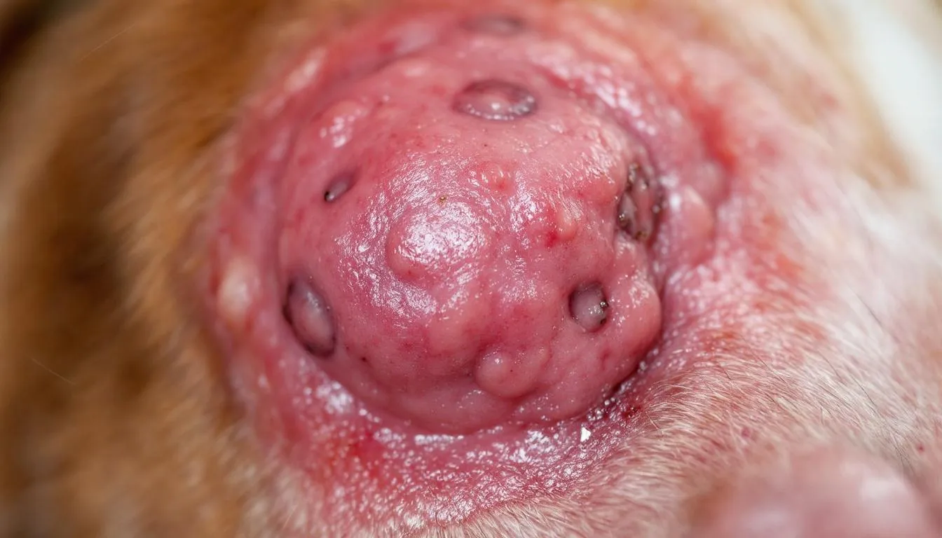
Deep pyoderma represents the most severe form, with bacterial infection extending into the dermis and subcutaneous tissue following follicular rupture. This condition often produces systemic signs including fever, malaise, and lymphadenopathy, requiring immediate veterinary intervention.
The clinical presentation includes nodules, draining tracts, ulcerations, and hemorrhagic exudate, often accompanied by significant pain that may limit the dog’s mobility and appetite. Deep infections commonly affect the muzzle, pressure points such as elbows and hocks, and interdigital spaces where constant pressure and moisture create predisposing conditions.
German Shepherd Dogs show particular predisposition to deep pyoderma, often developing extensive lesions on the caudal thighs and ventral abdomen. These cases frequently require prolonged systemic antimicrobial therapy and may result in permanent scarring or pigmentation changes. Systemic therapy is the primary approach for managing deep pyoderma, especially when resistant strains of bacteria are involved, as these can complicate management and necessitate alternative or adjunctive treatment strategies.
The progression from superficial to deep pyoderma can occur when underlying conditions remain uncontrolled or when inappropriate treatment allows bacterial proliferation to overwhelm local immune defenses. Once deep infection establishes, the treatment becomes significantly more challenging and expensive.

Clinical Signs and Symptoms
Recognition of pyoderma requires familiarity with both primary and secondary lesions that develop throughout the infection process. Primary lesions represent the initial bacterial infection, while secondary changes result from self-trauma, chronicity, or treatment effects.
Staphylococcal infections are a frequent underlying cause of these clinical signs, with Staphylococcus pseudintermedius and Staphylococcus aureus commonly implicated in the development of lesions on the skin surface.
Primary lesions include papules (small, solid elevations), pustules (pus-filled elevations), follicular casts (keratin plugs within hair follicles), and the pathognomonic epidermal collarettes. These lesions typically appear first in areas of reduced hair density or increased moisture retention.
Secondary changes develop as the infection progresses or becomes chronic, including crusts formed from dried exudate, scales representing abnormal keratinization, hyperpigmentation from chronic inflammation, and lichenification indicating skin thickening from prolonged irritation.
The intensity of pruritus varies significantly based on the underlying cause and infection depth. Dogs with atopic dermatitis-associated pyoderma often display intense itchy skin that leads to extensive self-trauma, while those with endocrine-related infections may show minimal scratching despite extensive lesions.
Systemic signs become prominent in deep pyoderma cases, with affected dogs displaying decreased appetite, lethargy, and elevated body temperature. Regional lymphadenopathy frequently accompanies deep infections, and severe cases may progress to cellulitis or sepsis requiring hospitalization.
The distribution pattern of skin lesions often provides clues to underlying causes. Ventral distribution suggests contact allergens or bacterial overgrowth in warm, moist areas, while dorsal lesions may indicate flea allergy or other ectoparasitic infections.
Underlying Causes and Risk Factors
Successful management of canine pyoderma requires identification and control of underlying causes that predispose dogs to bacterial skin infections. These predisposing factors can be broadly categorized into allergic, parasitic, endocrine, and immunosuppressive conditions.
Atopic dermatitis represents the most common underlying cause in companion animal infectious diseases, with the chronic inflammation and barrier dysfunction creating ideal conditions for bacterial colonization. The constant scratching and licking associated with canine atopic dermatitis introduce bacteria into damaged skin while disrupting the normal protective mechanisms.
Food allergies similarly contribute to recurrent superficial pyoderma through chronic inflammation and barrier compromise. Dogs with food sensitivities often develop pyoderma in characteristic locations such as the feet, face, and perianal areas, requiring both antimicrobial therapy and dietary management.
Parasitic infections, particularly demodicosis, create significant predisposition to secondary bacterial infections. Demodex mites damage hair follicles and suppress local immune responses, allowing bacterial overgrowth to establish. Flea allergies contribute through the intense pruritus and self-trauma that breach the skin barrier. Skin scrapings are commonly performed to identify mites and rule out parasitic causes of skin disease.
Endocrine disorders profoundly impact skin health and infection susceptibility. Hypothyroidism alters lipid metabolism and immune function, while hyperadrenocorticism (Cushing’s disease) suppresses inflammatory responses and delays wound healing. Diabetes mellitus provides elevated glucose levels that support bacterial growth while impairing immune cell function.
Immunosuppressive conditions, whether primary immunodeficiencies or secondary to medications such as corticosteroids, significantly increase the risk of developing resistant infections. Dogs receiving chemotherapy or other immunosuppressive treatments require careful monitoring for skin infections that may progress rapidly.
Anatomical factors also contribute substantially to pyoderma development. Skin folds create warm, moist environments with poor air circulation, while obesity exacerbates fold dermatitis and creates additional pressure points. Breeds with excessive wrinkling, such as Shar-Peis and English Bulldogs, face lifelong predisposition to recurrent skin fold pyoderma.
As part of the differential diagnosis, fungal cultures may be performed to exclude fungal skin diseases.
Diagnostic Approach
Accurate diagnosis of canine pyoderma follows a systematic three-step process designed to confirm bacterial involvement, identify causative organisms, and determine appropriate treatment protocols while minimizing inappropriate antimicrobial use.
The initial step involves thorough skin examination following proper hair clipping and debris removal to visualize all lesions clearly. This examination should document lesion type, distribution, and severity while identifying any systemic signs that might indicate deep infection or sepsis.
Skin cytology represents the mandatory second step, providing rapid confirmation of bacterial involvement with 93% diagnostic sensitivity when properly performed. This cost-effective diagnostic tool can be completed in most veterinary practices within minutes, making it an essential component of routine dermatological evaluation.
The third step, bacterial culture and susceptibility testing, is reserved for specific indications including deep pyoderma, treatment failures, or suspected antimicrobial resistance. Fungal culture should also be considered to confirm or rule out dermatophyte infections, especially when clinical signs are ambiguous or when other tests suggest a possible fungal etiology. This selective approach balances diagnostic accuracy with cost considerations while supporting antimicrobial stewardship principles.
In difficult or non-responsive cases, a skin biopsy may be warranted to obtain histopathological evaluation and clarify the diagnosis when clinical findings and initial tests are inconclusive.
Cytological Examination
Proper sample collection techniques ensure accurate cytological interpretation and appropriate treatment decisions. Impression smears from moist lesions, tape strips from dry areas, and fine-needle aspirates from deep nodules each provide specific information about the infection type and severity.
Microscopic examination reveals characteristic findings including neutrophils, cocci bacteria arranged in clusters or chains, and various inflammatory changes that confirm bacterial involvement. The presence of intracellular bacteria indicates active infection requiring antimicrobial intervention.
Differentiation from non-infectious skin diseases relies on identifying bacterial organisms and inflammatory cells while ruling out other pathogens such as yeasts, parasites, or neoplastic cells. Mixed infections involving both bacteria and Malassezia yeasts commonly occur, requiring combination therapy approaches.
The cost-effectiveness and rapid availability of cytology make it the preferred initial diagnostic tool, with most veterinary clinics capable of performing and interpreting results within their standard workflow. This accessibility supports evidence-based treatment decisions without delaying appropriate therapy.
Bacterial Culture and Susceptibility Testing
Indications for bacterial culture include deep pyoderma, recurrent infections despite appropriate therapy, previous treatment failures, or clinical suspicion of antimicrobial resistance based on patient history or regional resistance patterns.
Proper sampling techniques vary by lesion type, with intact pustules providing the most reliable samples when available. For crusted or dry lesions, the area should be cleaned and sampled from beneath the crust where viable bacteria are most likely to be present.
Interpretation of culture results requires understanding normal skin flora and distinguishing pathogenic organisms from contaminants. Mixed cultures often reflect surface contamination rather than true polymicrobial infection, emphasizing the importance of proper sample collection and handling. Antimicrobial susceptibility testing is essential for guiding targeted therapy, as it helps select the most effective drugs, especially in cases involving resistant bacteria.
Cost considerations and laboratory selection criteria include turnaround time, susceptibility testing panels, and communication of results. Many commercial laboratories provide rapid turnaround for routine cases while offering expanded testing for complex or resistant infections. Adjunctive treatment can also play a valuable role in improving outcomes and reducing the need for systemic antibiotic use.
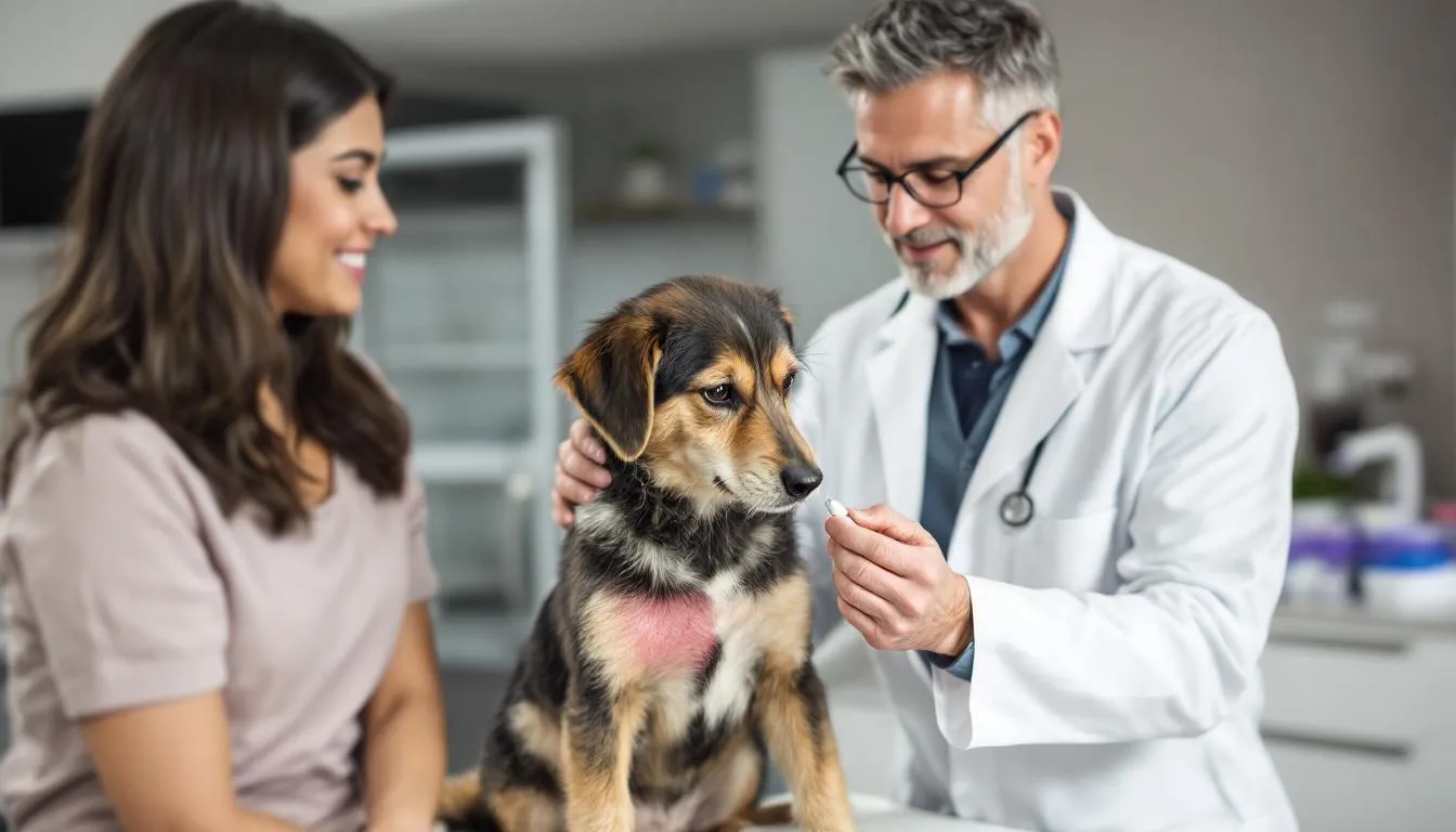
Treatment Protocols
Treatment selection depends on accurate assessment of infection depth and severity, with topical antimicrobial therapy preferred for surface and superficial pyoderma, while systemic antibiotics are reserved for deep infections or cases where topical therapy has failed. In certain cases, such as localized lesions or infections caused by meticillin-resistant staphylococci, topical antibiotics like fusidic acid or mupirocin may be used when antiseptics are insufficient, based on cytology results and evidence of effectiveness.
The paradigm shift toward antimicrobial stewardship emphasizes using the least intensive effective therapy to minimize resistance development while achieving clinical cure. This approach requires careful evaluation of each case and willingness to adjust treatment based on response and culture results. The veterinary clinic plays a crucial role in performing diagnostic tests, such as physical exams, cultures, and sensitivity testing, to guide appropriate therapy and ensure infection control.
Concurrent management of underlying predisposing conditions remains essential for long-term success, as failure to address these factors often results in treatment failure or rapid recurrence despite appropriate antimicrobial therapy.
Topical Antimicrobial Therapy
First-line topical agents include chlorhexidine, benzoyl peroxide, and ethyl lactate, each offering unique advantages for different clinical situations. Chlorhexidine provides broad-spectrum antimicrobial activity with excellent safety profiles, while benzoyl peroxide offers additional keratolytic and follicular flushing properties.
Product formulations range from medicated shampoos for whole-body treatment to mousses, wipes, and leave-on treatments for localized application. The selection depends on lesion location, dog size, owner compliance factors, and specific product characteristics.
Application techniques significantly impact treatment efficacy, with proper contact time, frequency, and coverage being critical for success. Shampoos typically require 10-15 minute contact time, while leave-on products should be applied to completely dry skin for optimal penetration.
The advantages of topical treatment include reduced systemic exposure, decreased resistance pressure, localized high concentrations, and support for antimicrobial stewardship principles. These benefits make topical therapy the preferred first-line approach for most surface and superficial infections.
Systemic Antibiotic Therapy
First-choice systemic antimicrobials for empirical treatment include clindamycin (11-33 mg/kg twice daily), cefalexin (22-35 mg/kg twice daily), and amoxicillin-clavulanate (13.75-25 mg/kg twice daily), selected based on spectrum, penetration, and resistance patterns.
Second-choice options such as potentiated sulfonamides, chloramphenicol, and macrolides are reserved for cases with contraindications to first-line drugs or confirmed resistance patterns. These alternatives often require more frequent monitoring due to potential adverse effects.
Treatment duration follows established guidelines with 3-4 weeks for superficial pyoderma and potentially months for deep infections, with therapy continuing 7-14 days beyond apparent clinical cure to prevent recurrence and minimize resistance development.
Monitoring protocols include regular re-evaluation at 2-week intervals for superficial cases and weekly for deep infections, with adjustments based on clinical response, adverse effects, and culture results when available.
Antibiotic Resistance Management
Methicillin-resistant Staphylococcus pseudintermedius (MRSP) represents a growing concern in veterinary medicine, with prevalence rates of 5-15% reported in some North American and European studies, particularly in referral populations with repeated antibiotic exposure.
The implications of resistant infections extend beyond individual patient care to include increased treatment costs, longer treatment durations, limited therapeutic options, and potential zoonotic risks. These factors emphasize the importance of resistance prevention through appropriate antimicrobial use.
Culture-guided therapy becomes essential for resistant organisms and multidrug-resistant infections, as empirical treatment often fails and may worsen resistance patterns. Susceptibility testing should include both routine antimicrobials and reserve drugs based on clinical indication.
Reserved antimicrobials such as fluoroquinolones, third-generation cephalosporins, and newer agents should only be used based on culture results and specialist consultation, following guidelines from the antimicrobial guidelines working group to preserve their effectiveness.
Infection control measures in veterinary hospitals include proper hand hygiene, equipment disinfection, and isolation protocols for known resistant cases. These measures prevent cross-contamination and reduce environmental persistence of resistant organisms.
Role of the International Society for Companion Animal Infectious Diseases (ISCAID)
The International Society for Companion Animal Infectious Diseases (ISCAID) stands at the forefront of advancing best practices in the diagnosis, treatment, and prevention of infectious diseases in dogs and cats, including pyoderma in dogs. As a leading authority in veterinary medicine, ISCAID is dedicated to promoting responsible antimicrobial therapy and combating the growing threat of antimicrobial resistance in companion animal infectious diseases.
ISCAID’s antimicrobial guidelines working group develops and regularly updates evidence-based recommendations for managing bacterial skin infections, such as superficial pyoderma and recurrent infections. These guidelines are grounded in the latest clinical research and expert consensus, ensuring that veterinarians have access to the most current and effective strategies for treating pyoderma in dogs.
A cornerstone of ISCAID’s approach is the emphasis on identifying and addressing the underlying cause of skin disease—whether it be allergies, anatomical factors like skin folds, or immune system dysfunction. By focusing on the root of the problem, ISCAID aims to reduce the risk of recurrent infections and improve long-term outcomes for affected dogs.
The society advocates for a stepwise diagnostic process, beginning with a thorough assessment of clinical signs, followed by skin cytology to confirm bacterial involvement. When indicated, ISCAID recommends bacterial culture and susceptibility testing to guide targeted antimicrobial therapy, especially in cases of treatment failure or suspected antimicrobial resistance. This approach helps ensure that systemic antibiotics are used judiciously and only when necessary, preserving their effectiveness for more severe or deep infections.
ISCAID also strongly supports the use of topical therapy—such as medicated shampoos and antimicrobial creams—as the first-line treatment for superficial pyoderma. By prioritizing topical treatments, veterinarians can achieve clinical cure while minimizing the need for systemic antibiotics and reducing the risk of resistance development.
Beyond clinical recommendations, ISCAID provides valuable resources and ongoing education for veterinary professionals, helping them stay informed about emerging trends and best practices in companion animal infectious diseases. By fostering a culture of antimicrobial stewardship and evidence-based care, ISCAID not only improves the health and well-being of dogs with pyoderma but also contributes to the global effort to safeguard both animal and public health from the dangers of antimicrobial resistance.
Prevention and Long-term Management
Identification and control of underlying primary causes represents the cornerstone of successful long-term management, as dogs requiring systemic antimicrobials more than once yearly need intensified investigation for predisposing conditions.
Maintenance topical antiseptic therapy using chlorhexidine-based products 2-3 times weekly can effectively prevent recurrence in dogs with chronic predisposing conditions while minimizing systemic antibiotic exposure.
Management of canine atopic dermatitis through anti-inflammatory medications, allergen avoidance, and immunomodulatory treatments significantly reduces the frequency and severity of secondary bacterial infections. This integrated approach addresses both the underlying inflammation and its infectious complications.
Regular monitoring schedules should be established for dogs with chronic conditions, with owner education on recognizing early signs enabling prompt intervention before infections become severe or deep.
Recurrence Prevention Strategies
Proactive topical chlorhexidine therapy applied 2-3 times weekly to previously affected areas can maintain the skin barrier and prevent bacterial recolonization in high-risk dogs. This approach proves particularly valuable for dogs with anatomical predispositions or chronic allergic conditions.
Allergen avoidance and environmental management strategies include identification and elimination of contact allergens, flea prevention protocols, and dietary management for food-allergic dogs. These measures address upstream causes that initiate the inflammatory cascade leading to skin barrier compromise.
Immunomodulatory treatments such as allergen-specific immunotherapy, cyclosporine, or oclacitinib can reduce the inflammatory burden in allergic dogs, thereby decreasing their susceptibility to secondary bacterial infections.
Owner education programs focusing on early recognition of clinical signs, proper hygiene practices, and when to seek veterinary care enable timely intervention that prevents progression from mild surface infections to severe deep pyoderma.
Prognosis and Complications
Excellent outcomes can be achieved with appropriate therapy and underlying cause management, with most surface and superficial infections resolving completely within 3-4 weeks of appropriate treatment initiation.
However, several potential complications can impact prognosis, including the development of antimicrobial resistance, treatment failures due to inadequate duration or inappropriate drug selection, and chronic recurrence when underlying conditions remain uncontrolled.
Deep pyoderma carries increased risk of complications including cellulitis, sepsis, and systemic involvement that may require hospitalization and intensive care. These cases often result in permanent scarring, pigmentation changes, or functional impairment.
Long-term management requirements for dogs with chronic underlying conditions may include lifelong maintenance therapy, regular monitoring, and periodic adjustments based on disease progression or treatment response.

Zoonotic Considerations
The transmission risk of staphylococcus pseudintermedius from dogs to humans remains low, as this organism rarely causes clinical disease in people with normal immune function. However, direct contact with infected exudate should be minimized through proper hygiene practices.
Higher concern exists for cases involving methicillin-resistant staphylococcus aureus (MRSA), which can cause serious infections in humans, particularly immunocompromised individuals. These cases require enhanced infection control measures and potential isolation protocols.
Hygiene recommendations for households with affected dogs include regular hand washing after contact, avoiding direct contact with infected lesions, and seeking medical attention if skin infections develop in family members, particularly those with compromised immune systems.
Infection control protocols during veterinary visits should include proper hand hygiene, examination glove use, and equipment disinfection to prevent cross-contamination between patients and transmission to veterinary staff.
FAQ
How long does it take for pyoderma to heal in dogs?
Superficial pyoderma typically requires 3-4 weeks of treatment, while deep pyoderma may need several months of therapy. Treatment should continue 7-14 days after the skin appears normal to prevent recurrence and antibiotic resistance. The healing time depends on the infection depth, underlying causes, and how quickly appropriate treatment begins.
Can dogs with pyoderma infect other pets or humans?
Transmission between dogs is uncommon, and human infection from canine pyoderma is rare since Staphylococcus pseudintermedius rarely causes disease in humans. However, proper hygiene should be maintained, especially around immunocompromised individuals. Cases involving MRSA pose higher zoonotic risk and require enhanced precautions.
Why does my dog keep getting pyoderma despite antibiotic treatment?
Recurrent pyoderma indicates an underlying condition that hasn’t been identified or properly managed, such as allergies, endocrine disorders, or parasites. Dogs requiring systemic antimicrobials more than once yearly need intensified investigation for primary causes. Without addressing these underlying factors, infections will continue to recur despite appropriate antibiotic therapy.
Are there alternatives to antibiotics for treating pyoderma?
Topical antimicrobial therapy using chlorhexidine, benzoyl peroxide, or ethyl lactate can effectively treat surface and superficial pyoderma without systemic antibiotics. Autogenous bacterins and immunomodulatory treatments may help reduce antibiotic dependence in recurrent cases. These alternatives support antimicrobial stewardship while maintaining clinical efficacy.
What should I do if my dog’s pyoderma doesn’t improve with treatment?
Contact your veterinarian immediately if no improvement is seen within the first few days of treatment. This may indicate antibiotic resistance, incorrect diagnosis, or inadequate management of underlying causes, requiring bacterial culture and treatment adjustment. Early intervention prevents progression to deeper, more serious infections that are harder to treat.


