


Key Takeaways
Canine eye ulcers are painful wounds on the cornea that require immediate veterinary attention to prevent vision loss
- Canine eye ulcers are painful wounds on the cornea that require immediate veterinary attention to prevent vision loss
- The cause of canine eye ulcers is most commonly trauma, especially in brachycephalic breeds like pugs and bulldogs
- Fluorescein stain testing is the primary diagnostic tool that makes ulcers glow green under special light
- Treatment ranges from antibiotic drops for superficial ulcers to emergency surgery for deep ulcers affecting the stroma
- With proper treatment, most corneal ulcers heal within 3-10 days, but complicated cases may require specialist referral
The cause of canine eye ulcers is most commonly trauma, especially in brachycephalic breeds like pugs and bulldogs
Fluorescein stain testing is the primary diagnostic tool that makes ulcers glow green under special light
Treatment ranges from antibiotic drops for superficial ulcers to emergency surgery for deep ulcers affecting the stroma
With proper treatment, most corneal ulcers heal within 3-10 days, but complicated cases may require specialist referral
When your dog suddenly starts squinting, pawing at their eye, or showing signs of intense pain around their face, you could be witnessing the early stages of a corneal ulcer. This serious eye condition affects thousands of dogs annually and demands immediate veterinary care to preserve vision and prevent devastating complications.
A canine eye ulcer represents an open wound on the cornea—the clear, protective dome covering the front of your dog’s eye. Unlike minor irritations that resolve on their own, corneal ulcers create a pathway for bacteria and other harmful agents to penetrate deeper eye structures, potentially leading to vision loss or even eye rupture if left untreated.
Understanding the signs, causes, and treatment options for ulcers in dogs empowers you to act swiftly when your pet needs help most. This comprehensive guide covers everything from recognizing early symptoms to navigating complex treatment decisions, ensuring you’re prepared to protect your dog’s precious eyesight.
Understanding the Canine Cornea
The cornea serves as your dog’s primary protective barrier for inner eye structures, functioning like a transparent shield that allows light to pass through while keeping harmful elements out. This remarkable layer of the eye measures approximately half the width of a dime but performs multiple critical functions that make vision possible.
The corneal structure consists of three distinct layers, each playing a vital role in maintaining eye health. The outermost corneal epithelium acts as the first line of defense, composed of highly specialized skin cells that constantly regenerate to maintain a smooth, clear surface. Beneath this protective coating lies the corneal stroma, which represents the main supportive tissue and comprises about 90% of the cornea’s thickness. The innermost very thin layer, called Descemet’s membrane, provides structural integrity and prevents fluid from entering the cornea.
Unlike most body tissues, the cornea lacks its own blood supply, instead receiving nutrients from tears and the fluid inside the eye. This unique characteristic allows the cornea to remain transparent for clear vision but also makes healing more challenging when injuries occur. The corneal surface contains an extensive network of nerve endings, making any damage to this layer of the eye extremely painful for your dog.
The cornea’s smooth surface requires constant moisture from tear production to function properly. When tear film becomes insufficient or of poor quality, the corneal epithelium becomes vulnerable to breakdown and subsequent ulceration. This explains why dogs with dry eye syndrome frequently develop corneal ulcers as a secondary complication. Corneal dystrophy is another condition that can cause corneal opacity in dogs; unlike scarring from ulcers, corneal dystrophy is typically genetic or metabolic in origin and not related to injury or tear film deficiency.
What Is a Deep Canine Corneal Ulcer?
A corneal ulcer, medically termed ulcerative keratitis, represents an erosion or open wound that develops when the protective surface epithelium of the cornea becomes damaged or destroyed. Unlike a simple corneal abrasion, which affects only the surface epithelium, a true ulcer extends deeper into the corneal tissue and requires more aggressive medical intervention. Corneal erosion refers specifically to a superficial epithelial injury; if not managed, it can progress to a deeper corneal ulcer, impacting corneal transparency and potentially leading to more serious complications.
Veterinarians classify corneal ulcers based on their depth and complexity. A superficial corneal ulcer involves only the outermost epithelial layer and typically heals within several days with proper treatment. These surface injuries, while painful, generally carry an excellent prognosis when managed promptly and appropriately.
Deep corneal ulcers present a much more serious situation, as they extend into the corneal stroma and threaten the structural integrity of the entire eye. When an ulcer extends deep enough to expose the thin layer beneath the stroma, called Descemet’s membrane, the condition becomes a veterinary emergency requiring immediate surgical intervention to prevent eye rupture.
The most severe form occurs when descemet’s membrane ruptures, leading to complete corneal perforation. This catastrophic complication results in immediate loss of the eye’s internal contents and permanent blindness in the affected eye. Such cases often require emergency eye removal to prevent life-threatening complications from spreading infection.
Indolent corneal ulcers represent a frustrating subset of superficial ulcers that fail to heal despite appropriate treatment. These chronic wounds typically affect older dogs and may persist for weeks or months without surgical intervention. The healing process stalls because the surrounding epithelial cells cannot properly attach to the underlying tissue, creating a non-healing wound that requires specialized treatment approaches. Epithelial dystrophy, an inherited condition affecting the corneal structure, can predispose certain breeds to chronic or recurrent corneal ulceration and dystrophic changes.
Common Causes of Canine Eye Ulcers in Dogs
Trauma stands as the primary culprit behind most canine eye ulcers, with everyday activities posing unexpected risks to your dog’s corneal surface. Blunt force injuries from running into furniture, getting hit by toys, or colliding with other dogs can instantly damage the delicate corneal epithelium. Superficial corneal ulcers resulting from these incidents are considered a much less serious injury compared to deep ulcers, as they affect only the surface epithelium, but they still require prompt veterinary care. Cat scratches represent particularly dangerous trauma sources, as feline claws carry bacteria that can rapidly transform a simple scratch into a severe bacterial infection.
Chemical irritants frequently cause corneal ulceration when household products accidentally contact your dog’s eyes. Shampoo during bath time, cleaning products, drywall dust during home renovations, and certain plant materials can chemically burn the corneal surface. Even seemingly harmless activities like applying flea treatments or ear medications can result in chemical trauma if these substances accidentally drip into the eyes.
Brachycephalic breeds—including pugs, bulldogs, Boston terriers, and French bulldogs—face significantly higher risks for developing eye ulcers due to their anatomical features. These dogs possess prominent eyes that protrude beyond the protective socket, making the corneal surface more vulnerable to environmental hazards and trauma. Additionally, their facial structure often leads to inadequate eyelid closure, causing chronic dryness and increased ulceration risk.
Underlying diseases frequently predispose dogs to corneal ulceration. Dry eye syndrome, or keratoconjunctivitis sicca, reduces tear production and compromises the corneal surface’s natural protective mechanisms. Eyelid abnormalities such as entropion (inward rolling eyelids) or abnormal eyelashes can create constant irritation that eventually leads to ulceration. Endocrine disorders like diabetes mellitus can impair the healing process and increase susceptibility to bacterial infections. In cases where ulcers do not heal as expected, it is crucial to identify and address the underlying cause, such as dry eye, eyelash abnormalities, or entropion, to ensure effective treatment and resolution.
Foreign body injury occurs when small particles become trapped under the eyelids, creating ongoing mechanical trauma to the corneal surface. Common culprits include grass seeds, sand, dust particles, and loose hairs that work their way under the third eyelid. Self trauma from pawing at already irritated eyes often worsens minor surface injuries into full ulcers, highlighting the importance of addressing eye discomfort promptly.
Bacterial infections can either cause primary ulceration or complicate existing corneal injuries. When bacteria release substances that break down corneal tissue, they create rapidly expanding wounds called melting ulcers that can perforate the eye within hours. This represents one of the most dangerous complications of corneal ulceration and requires immediate emergency veterinary care.
Recognizing Symptoms of Canine Eye Ulcers
The most obvious indicator of a canine eye ulcer is intense pain that manifests through distinctive behavioral changes. Dogs with corneal ulcers typically exhibit persistent squinting, keeping the affected eye partially or completely closed to minimize discomfort. This protective response, called blepharospasm, represents your dog’s natural attempt to shield the damaged corneal surface from further irritation.
Pawing at the affected eye or rubbing the face against furniture signals significant pain and discomfort. Dogs may also shake their heads frequently or show reluctance to have their face touched. These behaviors can worsen the underlying condition by introducing bacteria from dirty paws or causing additional mechanical trauma to the already compromised corneal surface.
Visual changes in the eye itself provide important diagnostic clues about ulcer severity and depth. The corneal surface may appear cloudy, hazy, or develop a blue-white opacity that wasn’t present before. This cloudiness, called corneal edema, occurs when the damaged epithelium allows fluid to accumulate within the corneal layers. Deeper ulcers may create visible depressions or craters in the corneal surface that can be seen with careful observation.
Discharge from the affected eye varies depending on whether bacterial infection has developed. Clear, watery tearing represents the eye’s initial response to corneal damage, while yellow or green discharge indicates bacterial involvement requiring immediate antibiotic treatment. Bloody discharge suggests more severe tissue damage or possible trauma to deeper eye structures.
The third eyelid may become elevated and more visible as part of the body’s protective response to ocular pain. This pink membrane, normally hidden in the inner corner of the eye, may partially cover the eye surface when significant pain or inflammation develops. While not specific to ulcers, third eyelid elevation combined with other symptoms warrants prompt veterinary evaluation.
Systemic signs of discomfort may include loss of appetite, lethargy, or general behavioral changes as your dog copes with ongoing pain. Some dogs become photophobic, seeking dark areas and avoiding bright lights that intensify their discomfort. These behavioral changes often prompt owners to seek veterinary care even when eye-specific symptoms might be subtle.
Emergency signs requiring immediate veterinary attention include sudden onset of severe pain, visible holes or depressions in the corneal surface, or any indication that the eye contents are leaking. A soft, collapsed appearance of the eyeball or obvious fluid drainage signals possible corneal perforation—a sight-threatening emergency requiring immediate surgical intervention.
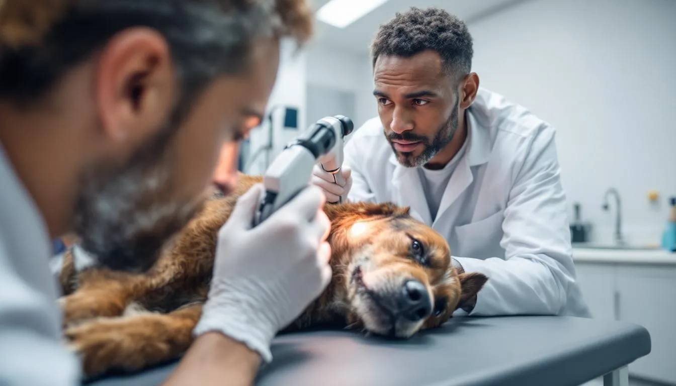
Veterinary Ophthalmologist Diagnosis of Eye Ulcers
Veterinary diagnosis of corneal ulcers begins with a comprehensive ophthalmic examination that evaluates both the obvious symptoms and subtle signs that might indicate underlying complications. Your veterinarian will first assess your dog’s level of pain and discomfort, noting behavioral indicators like squinting, head shaking, or reluctance to open the affected eye.
The fluorescein stain test serves as the gold standard for diagnosing corneal ulcers and represents one of the most important diagnostic tools in veterinary ophthalmology. During this procedure, your veterinarian places a small drop of orange fluorescein dye onto the eye surface, then rinses it away with saline solution. Healthy corneal epithelium repels the dye, but areas where the epithelium has been lost will retain the fluorescein, creating a bright green fluorescence under blue light examination.
This fluorescein staining pattern reveals not only the presence of an ulcer but also its size, shape, and approximate depth. Linear patterns might suggest foreign body trauma, while irregular or expanding patterns could indicate bacterial involvement. The intensity of fluorescein uptake helps determine whether the ulcer affects only the superficial epithelium or extends into deeper corneal layers.
Additional diagnostic tests help identify underlying causes and potential complications. The Schirmer tear test measures tear production by placing small paper strips in the lower eyelid for one minute. Reduced tear production indicates dry eye syndrome, which requires specific treatment to prevent ulcer recurrence. Tonometry measures intraocular pressure to rule out glaucoma, which can complicate ulcer healing and cause additional pain.
When bacterial infection is suspected, your veterinarian may collect samples for bacterial culture and sensitivity testing. This involves gently swabbing the ulcer surface to identify specific bacterial organisms and determine which antibiotics will be most effective. Culture results typically take 2-3 days but provide crucial information for treating resistant or complicated infections.
Classification of ulcers as simple versus complicated determines the appropriate treatment approach and prognosis. Simple ulcers are superficial, show signs of healing within 2-3 days of treatment, and lack evidence of infection. Complicated ulcers involve deeper corneal layers, show signs of bacterial infection, fail to respond to initial treatment, or occur in high-risk breeds prone to healing problems.
Advanced diagnostic techniques may be necessary for complicated cases. Corneal cytology examines cells from the ulcer surface under a microscope to identify inflammatory cells, bacteria, or abnormal cellular changes. In severe cases, referral to a veterinary ophthalmologist allows access to specialized equipment like slit-lamp biomicroscopy for detailed examination of corneal layers and assessment of potential surgical needs.
Treatment Options for Canine Eye Ulcers
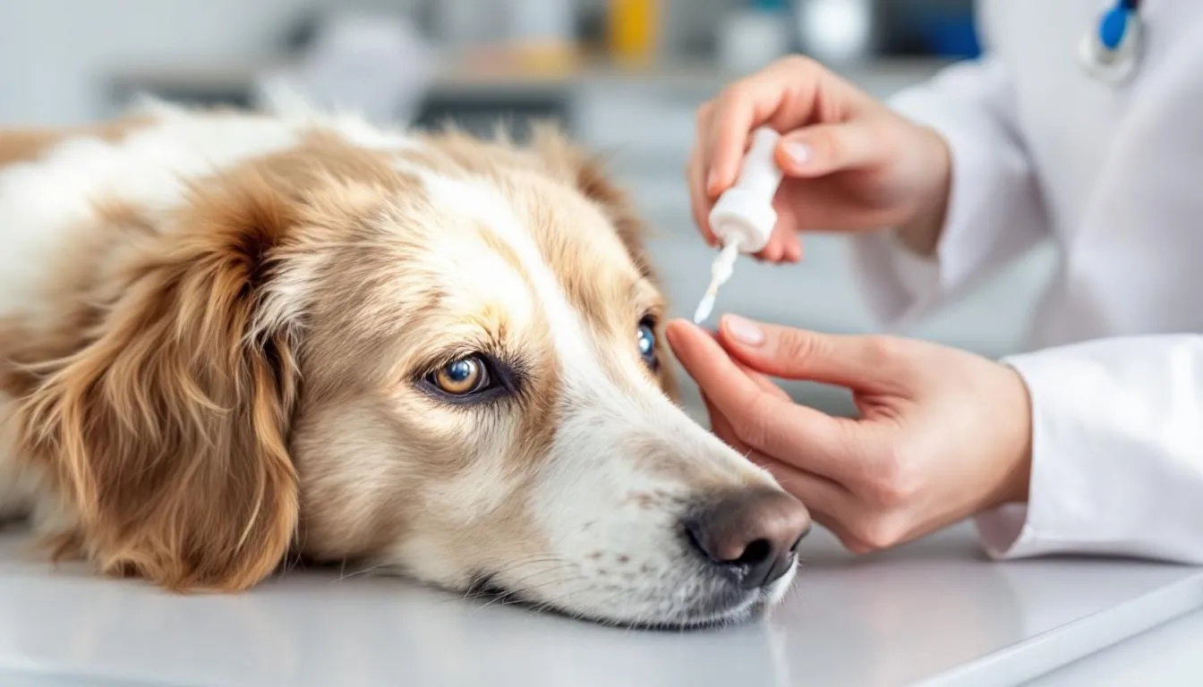
Treatment of uncomplicated superficial corneal ulcers focuses on preventing bacterial infection, managing pain, and creating optimal conditions for natural healing. Broad-spectrum topical antibiotic drops or ointments form the cornerstone of medical therapy, typically applied 3-6 times daily depending on the specific medication and ulcer severity. Common choices include antibiotic eye drops containing neomycin, polymyxin B, or fluoroquinolones that effectively target the most common bacterial contaminants.
Medical Therapy for Simple Ulcers
Pain management requires a multi-modal approach combining topical and systemic medications. Oral pain medications such as carprofen or gabapentin help manage the significant pain associated with corneal ulceration, while topical anesthetics may be used once during initial examination but are never prescribed for home use due to their potential to delay healing and mask worsening symptoms.
Atropine drops serve a dual purpose in ulcer treatment by dilating the pupil and reducing painful muscle spasms within the eye. This medication helps relieve discomfort while preventing secondary complications like glaucoma that can develop from chronic inflammation. The pupil dilation effect may last several days, causing temporary light sensitivity that resolves as treatment progresses.
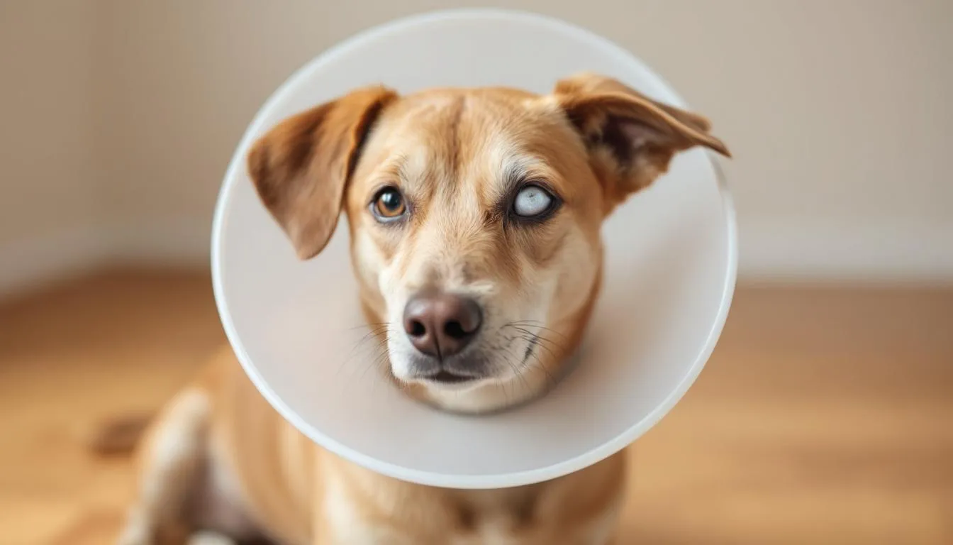
An Elizabethan collar represents perhaps the most critical component of successful ulcer treatment, preventing self trauma that can worsen the ulcer or introduce bacteria from dirty paws. Many treatment failures result from inadequate protection against pawing or rubbing, making collar compliance essential throughout the entire healing period. The collar should remain in place 24 hours daily until your veterinarian confirms complete healing through repeat fluorescein staining.
In select cases of uncomplicated ulcers, therapeutic contact lenses may provide additional protection and comfort. These specialized lenses act as a bandage, protecting the healing epithelium while allowing medication penetration. Contact lens placement requires specific expertise and regular monitoring to ensure proper fit and prevent complications.
The typical healing timeline for simple superficial ulcers ranges from 3-7 days with appropriate treatment and owner compliance. However, healing rates vary based on ulcer size, your dog’s overall health, and underlying factors like tear production adequacy. Most dogs show significant improvement within 48-72 hours of starting treatment, with decreased pain and squinting as early indicators of healing progress.
Advanced Treatment for Complicated Ulcers
Complicated corneal ulcers require more aggressive treatment approaches to address deeper tissue involvement, bacterial infection, or healing failure. These cases often necessitate referral to a veterinary ophthalmologist who possesses specialized training and equipment for advanced corneal procedures.
Intensive antibiotic therapy becomes necessary when culture results reveal specific bacterial organisms or when clinical signs suggest deep infection. Potent topical antibiotics may be applied every 1-2 hours initially, with frequency gradually reduced as the infection responds. Some cases require fortified antibiotic preparations specially compounded at higher concentrations than commercially available formulations.
Autologous serum eye drops, prepared from your dog’s own blood, provide growth factors and healing proteins that facilitate healing in stubborn cases. These drops are particularly valuable for treating indolent corneal ulcer cases that fail to respond to conventional therapy. The serum contains natural healing factors that promote epithelial cell attachment and proliferation.
Surgical debridement techniques address indolent ulcers that fail to heal due to poor epithelial adhesion. Diamond burr debridement uses a specialized rotating instrument to remove loose, non-adherent epithelial cells and create a fresh surface for healing. Grid keratotomy involves making superficial punctures in the cornea to stimulate new epithelial growth and improve cellular attachment.
Conjunctival grafts provide both structural support and blood supply for deep stromal ulcers that threaten corneal perforation. During this procedure, a small piece of conjunctiva (the pink tissue lining the eyelids) is surgically moved to cover the ulcer. The graft brings healing blood vessels to the avascular cornea while providing mechanical strength to prevent rupture.
Third eyelid flap procedures offer protection for severe ulcers by temporarily covering the corneal surface. The third eyelid is sutured across the eye opening, creating a protective barrier while healing occurs underneath. While this technique impairs vision temporarily, it can save eyes that might otherwise require removal.
Emergency surgical intervention becomes necessary when descemetocele formation or corneal perforation occurs. If descemet's membrane ruptures, fluid can leak from the eye, leading to potential collapse of the globe and severe damage. These sight-threatening complications require immediate surgical repair using techniques like tissue adhesives, amniotic membrane grafts, or emergency corneal transplantation. Success depends on rapid intervention before extensive eye damage develops.
Multiple eye drops and systemic pain medication may be required simultaneously in complicated cases, making owner compliance challenging but essential. Treatment protocols can involve 6-8 different medications applied at varying frequencies throughout the day, requiring dedicated commitment from pet owners to achieve successful outcomes.
Recovery and Follow-Up Care
The healing process for corneal ulcers requires careful monitoring and strict adherence to prescribed treatment protocols to ensure successful recovery and prevent complications. Understanding what to expect during recovery helps owners recognize normal healing versus warning signs that require immediate veterinary attention.
Superficial corneal ulcers typically begin showing improvement within 48-72 hours of starting appropriate treatment. Early positive signs include decreased squinting, reduced pawing at the eye, and improved willingness to open the affected eye. The corneal cloudiness may initially appear to worsen as inflammation develops, but this represents a normal part of the healing response rather than treatment failure.
Re-examination schedules vary based on ulcer severity and complexity, but most cases require follow-up appointments every 2-3 days initially. During these visits, your veterinarian will repeat the fluorescein stain test to assess healing progress and ensure the ulcer is not enlarging or deepening. Complete epithelial healing must be confirmed through negative fluorescein staining before discontinuing antibiotic therapy. It is essential that your veterinarian confirms the ulcer is completely healed before stopping any medications or removing protective devices.
Medication compliance represents the most critical factor determining treatment success. Antibiotic drops must be administered exactly as prescribed, even if your dog appears to be feeling better. Premature discontinuation of antibiotics can lead to treatment failure, bacterial resistance, or ulcer recurrence. Most uncomplicated ulcers require 7-14 days of antibiotic therapy, extending beyond the point of apparent clinical improvement.
The Elizabethan collar must remain in place throughout the entire treatment period and should only be removed under direct supervision for feeding or drinking. Many treatment failures result from owners removing the collar too early, allowing dogs to traumatize the healing corneal surface. The collar can be removed permanently only after your veterinarian confirms complete healing through repeat examination.
Monitoring for complications during recovery involves watching for specific warning signs that indicate treatment failure or developing problems. Worsening pain, increased squinting after initial improvement, changes in discharge color or amount, or development of new symptoms all warrant immediate veterinary contact. Progressive corneal cloudiness or the appearance of red blood vessels growing toward the ulcer may indicate either normal healing responses or developing complications requiring professional evaluation.
Environmental modifications during recovery help protect the healing eye from further trauma. Avoid dusty areas, prevent exposure to wind or air conditioning drafts, and supervise interactions with other pets that might accidentally injure the healing eye. Swimming or bathing should be avoided unless specifically approved by your veterinarian.
Long-term management focuses on addressing any underlying conditions that contributed to ulcer development. Dogs with dry eye syndrome require lifelong tear stimulant medications and regular monitoring. Brachycephalic breeds may benefit from environmental modifications and increased vigilance for early signs of ocular irritation.

Potential Complications and When to Seek Emergency Care
While most corneal ulcers heal successfully with appropriate treatment, several complications can develop that require immediate veterinary intervention to preserve vision and prevent more serious consequences. Understanding these potential problems helps owners recognize emergency situations and seek help before irreversible damage occurs.
Neovascularization, the growth of red blood vessels toward the ulcer site, represents a normal healing response that brings nutrients and immune cells to the avascular cornea. These vessels typically appear as thin red lines extending from the edge of the cornea toward the ulcer. While potentially alarming to owners, neovascularization usually indicates healthy healing progress rather than a complication requiring treatment modification.
Progressive ulcer deepening despite appropriate treatment suggests bacterial infection with organisms that produce enzymes capable of breaking down corneal tissue. These melting ulcers can rapidly progress from superficial wounds to full-thickness perforations within hours. Warning signs include increasing pain after initial improvement, rapidly expanding ulcer size, or development of a soft, gelatinous appearance to the corneal surface.
Descemetocele formation occurs when ulceration progresses through the entire corneal stroma, exposing the thin Descemet’s membrane as the only barrier preventing eye rupture. This condition appears as a clear, bubble-like protrusion from the corneal surface and represents a true ocular emergency requiring immediate surgical intervention. Any bulging or irregular corneal contour warrants emergency veterinary evaluation.
Corneal perforation results in immediate loss of the eye’s internal contents and permanent vision loss. Signs include a collapsed or soft eyeball, obvious fluid leakage from the eye, or a visible hole in the corneal surface. This devastating complication requires emergency surgery to seal the perforation and may necessitate eye removal if repair attempts fail.
Medication-related side effects occasionally develop during treatment and may require protocol adjustments. Topical antibiotics can cause increased sensitivity or irritation in some dogs, while atropine drops can cause decreased appetite due to their bitter taste when swallowed. Systemic pain medication may cause gastrointestinal upset or behavioral changes that concern owners.
Serious complications requiring immediate emergency veterinary care include sudden worsening of pain after initial improvement, visible changes in eye shape or size, obvious fluid leakage from the eye, or development of a white or yellow discharge with foul odor. Any of these signs suggests the development of severe infection, corneal perforation, or other sight-threatening complications.
Secondary glaucoma can develop when inflammation from ulceration blocks normal fluid drainage from the eye, causing dangerous increases in intraocular pressure. Signs include a hard, enlarged eyeball, severe pain, and rapid vision loss. This complication requires immediate treatment to prevent permanent damage to the optic nerve and retina.
Treatment failure, defined as lack of improvement or worsening after 3-5 days of appropriate therapy, indicates the need for treatment modification or specialist referral. Factors contributing to treatment failure include underlying dry eye, indolent ulcer formation, resistant bacterial infections, or concurrent conditions affecting healing ability.
Prevention Strategies for Dog Owners
Preventing canine eye ulcers requires a proactive approach that addresses both environmental hazards and breed-specific risk factors. Environmental safety measures form the foundation of ulcer prevention, starting with careful management of your dog’s surroundings to minimize trauma risks.
Remove or modify sharp objects and hazards from areas where your dog plays and exercises. Trim overgrown vegetation, especially plants with thorns or sharp leaves that could scratch the corneal surface during outdoor activities. Pay particular attention to rose bushes, blackberry canes, and ornamental grasses that commonly cause eye injuries. Consider protective barriers around garden areas or redirect walking routes to avoid high-risk vegetation.
Grooming precautions help prevent chemical and mechanical trauma during routine care activities. Protect your dog’s eyes during bathing by using tear-free shampoos and carefully controlling water flow to prevent soap contact with the corneal surface. Apply petroleum jelly around the eye area before bathing to create a protective barrier against shampoo and water. When trimming facial hair, use blunt-tipped scissors and work slowly to avoid accidental eye contact.
Brachycephalic breeds require special attention due to their anatomical predisposition to eye injuries. These dogs benefit from protective eyewear during outdoor activities in dusty or windy conditions. Consider doggy goggles or protective masks designed specifically for flat-faced breeds when hiking, riding in vehicles with open windows, or spending time in areas with airborne particles.
Regular eye examinations during routine veterinary visits enable early detection of predisposing conditions like dry eye syndrome or eyelid abnormalities. Many underlying causes of ulceration can be identified and treated before ulcer development occurs. Schedule comprehensive eye exams annually for healthy dogs, or more frequently for high-risk breeds or dogs with previous eye problems.
Early intervention for minor eye irritation prevents progression to full ulceration in many cases. Contact your veterinarian promptly if you notice increased tearing, mild squinting, or slight redness that persists beyond 24 hours. These subtle signs may indicate the beginning stages of corneal damage that can be reversed with prompt treatment.
Home monitoring involves daily observation of your dog’s eyes during routine interactions like feeding and grooming. Learn to recognize your dog’s normal eye appearance, including typical tear production and corneal clarity. Changes in pupil size, corneal transparency, or discharge production warrant veterinary evaluation even when pain signs are minimal.
Managing underlying conditions provides long-term protection against ulcer recurrence. Dogs with dry eye syndrome require lifelong tear production monitoring and medication adjustments as needed. Regular follow-up appointments ensure that tear stimulant medications remain effective and that corneal health is maintained over time.
Nutritional support for eye health includes ensuring adequate omega-3 fatty acid intake, which supports tear film quality and reduces inflammation. High-quality commercial diets typically provide sufficient nutrition for eye health, but supplements may be beneficial for dogs with chronic dry eye or recurrent eye problems. Discuss nutritional strategies with your veterinarian as part of a comprehensive eye health program.
FAQ
Can my dog go blind from an eye ulcer if left untreated?
Yes, untreated corneal ulcers can lead to permanent vision loss or complete blindness in the affected eye. Deep ulcers that progress to corneal perforation often result in irreversible damage to internal eye structures. Even superficial ulcers can cause significant corneal scarring that impairs vision if bacterial infection develops. However, with prompt veterinary treatment, most corneal ulcers heal completely without affecting your dog’s vision. The key is seeking care immediately when you notice symptoms like squinting, pawing at the eye, or discharge.
How long should I expect to give eye medications to my dog?
Most simple corneal ulcers require antibiotic treatment for 7-14 days, typically continuing for several days after the ulcer appears healed. Your veterinarian will determine the exact duration based on fluorescein stain test results that confirm complete epithelial healing. Complicated or deep ulcers may require several weeks of treatment, and some underlying conditions like dry eye syndrome need lifelong medication. Never discontinue eye medications early, even if your dog seems better—incomplete treatment can lead to ulcer recurrence or antibiotic resistance.
Is it normal for my dog’s eye to look worse before it gets better during treatment?
Yes, it’s common for the eye to appear more inflamed or cloudy during the first 2-3 days of treatment as the body’s healing response begins. The cornea may develop increased cloudiness, and you might notice blood vessels starting to grow toward the ulcer site. However, your dog should show decreased pain and squinting within 48-72 hours of starting treatment. If pain worsens after initial improvement, or if new symptoms develop like colored discharge or eye bulging, contact your veterinarian immediately as this may indicate complications.
What should I do if my dog won’t let me put the eye drops in?
Administering eye drops can be challenging, but proper technique and patience usually lead to success. Approach your dog calmly from behind or the side, gently lift the upper eyelid, and apply drops to the white part of the eye rather than directly onto the cornea. You can also place drops in the corner of the eye and allow them to distribute naturally when your dog blinks. If your dog remains too difficult to medicate, contact your veterinarian about alternative approaches like having the medications administered at the clinic or using different formulations that might be easier to apply.
Are certain dog breeds more likely to develop recurring eye ulcers?
Yes, brachycephalic breeds (pugs, bulldogs, Boston terriers, French bulldogs) have significantly higher rates of both initial ulcers and recurrence due to their prominent eyes and facial structure. These breeds often have inadequate eyelid closure and reduced tear film distribution that predisposes them to chronic corneal problems. Additionally, dogs with underlying conditions like dry eye syndrome, diabetes mellitus, or eyelid abnormalities face increased recurrence risk regardless of breed. Regular monitoring and management of predisposing factors can help reduce the likelihood of repeat ulcers in susceptible dogs.
Types of Corneal Ulcers in Dogs
Corneal ulcers in dogs are not all the same—understanding the specific type of ulcer your dog is facing can make a significant difference in both treatment and recovery. Veterinarians classify ulcers in dogs based on their depth, cause, and healing behavior, each with unique features and management strategies.
Superficial Corneal Ulcers: These are the most common type of corneal ulcers in dogs and involve only the outermost layer of the cornea, known as the epithelium. Superficial ulcers often result from minor trauma, such as a scratch or abrasion, and typically heal quickly with prompt veterinary care. While they can be very painful, superficial ulcers generally have an excellent prognosis when treated early and appropriately.
Deep Corneal Ulcers: When an ulcer extends beyond the surface epithelium into the underlying corneal stroma, it is classified as a deep corneal ulcer. These ulcers are much more serious, as they compromise the main supportive tissue of the cornea and increase the risk of complications like corneal perforation. Deep ulcers in dogs often require more intensive treatment, including stronger topical antibiotics, pain management, and sometimes surgical intervention to preserve the eye.
Indolent Corneal Ulcers (Non-Healing or Boxer Ulcers): Some ulcers, known as indolent corneal ulcers, fail to heal as expected despite appropriate therapy. These chronic ulcers are often seen in older dogs and certain breeds, such as Boxers, and are characterized by loose, non-adherent edges of the corneal epithelium. Indolent ulcers in dogs may persist for weeks or even months and often require specialized procedures like diamond burr debridement or grid keratotomy to stimulate proper healing.
Desmetoceles: A descemetocele forms when an ulcer has eroded through almost the entire thickness of the cornea, leaving only the very thin Descemet’s membrane intact. This type of ulcer is extremely fragile and represents an ophthalmic emergency, as the risk of corneal rupture is imminent. Immediate surgical intervention is usually necessary to save the affected eye.
Melting Ulcers: Melting ulcers are a rapidly progressing form of corneal ulceration where bacteria or inflammatory cells release enzymes that break down corneal tissue, causing the ulcer to appear soft and gelatinous. These ulcers in dogs can deteriorate quickly and require aggressive treatment with potent topical antibiotics and sometimes anti-collagenase medications to halt the destruction and facilitate healing.
Recognizing the type of corneal ulcer your dog has is essential for guiding treatment decisions and predicting recovery outcomes. If your veterinarian suspects a complicated or non-healing ulcer, referral to a veterinary ophthalmologist may be recommended to ensure your dog receives the most advanced care available.


