


Key Takeaways
- Canine conjunctivitis is inflammation of the conjunctiva (mucous membrane covering the eyeball and eyelids) causing red, swollen eyes with discharge
- Common causes include allergies, bacterial infections, viral infections, dry eye, and anatomical abnormalities requiring different treatment approaches
- Bacterial conjunctivitis typically resolves in 5-7 days with antibiotics, while viral forms may take 3-4 weeks to heal completely
- Early veterinary diagnosis is crucial as untreated conjunctivitis can lead to corneal scarring, chronic pain, and vision problems
- Most dogs recover well with proper treatment, though some conditions like allergies or dry eye may require lifelong management
Canine conjunctivitis is inflammation of the conjunctiva (mucous membrane covering the eyeball and eyelids) causing red, swollen eyes with discharge
Common causes include allergies, bacterial infections, viral infections, dry eye, and anatomical abnormalities requiring different treatment approaches
Bacterial conjunctivitis typically resolves in 5-7 days with antibiotics, while viral forms may take 3-4 weeks to heal completely
Early veterinary diagnosis is crucial as untreated conjunctivitis can lead to corneal scarring, chronic pain, and vision problems
Most dogs recover well with proper treatment, though some conditions like allergies or dry eye may require lifelong management
What is Canine Conjunctivitis?
Canine conjunctivitis represents one of the most frequently encountered ocular diseases in veterinary medicine. This condition involves inflammation of the conjunctiva, the transparent mucous membrane that covers the front surface of the eyeball and lines the inner aspects of the eyelids. The conjunctival epithelium is a nonkeratinized, stratified layer that serves as a barrier and plays a role in immune defense. This protective membrane serves as a crucial barrier against environmental irritants, bacteria, and other potential threats to the ocular surface. Resident lymphoid tissue contribute to the conjunctiva's rapid immune response to insults and inflammation.
Dogs possess a unique anatomical feature called the third eyelid or nictitating membrane, which is also composed of conjunctival tissue. The palpebral conjunctiva lines the inner surface of the eyelids and is involved in conjunctival inflammation, contributing to the clinical signs observed during a complete ophthalmic examination. Located at the inner corner of each eye, this additional protective structure can become involved in conjunctival inflammation, contributing to the clinical signs observed during a complete ophthalmic examination.
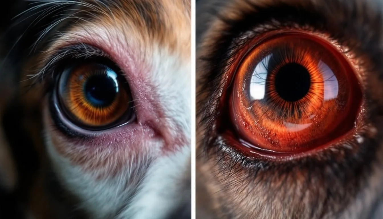
In healthy dogs, the conjunctiva appears pink and moist, though it may show pigmentation in certain breeds. The underlying connective tissue (substantia propria) beneath the epithelium contains blood vessels, nerves, and lymphoid tissue, supporting the conjunctiva's function and response to disease. During inflammatory episodes, this delicate tissue becomes swollen and turns bright pink or red, earning the condition its common name “pink eye.” The severity of conjunctival hyperemia and swelling can vary significantly depending on the underlying cause and duration of the condition.
Common Symptoms of Conjunctivitis in Dogs
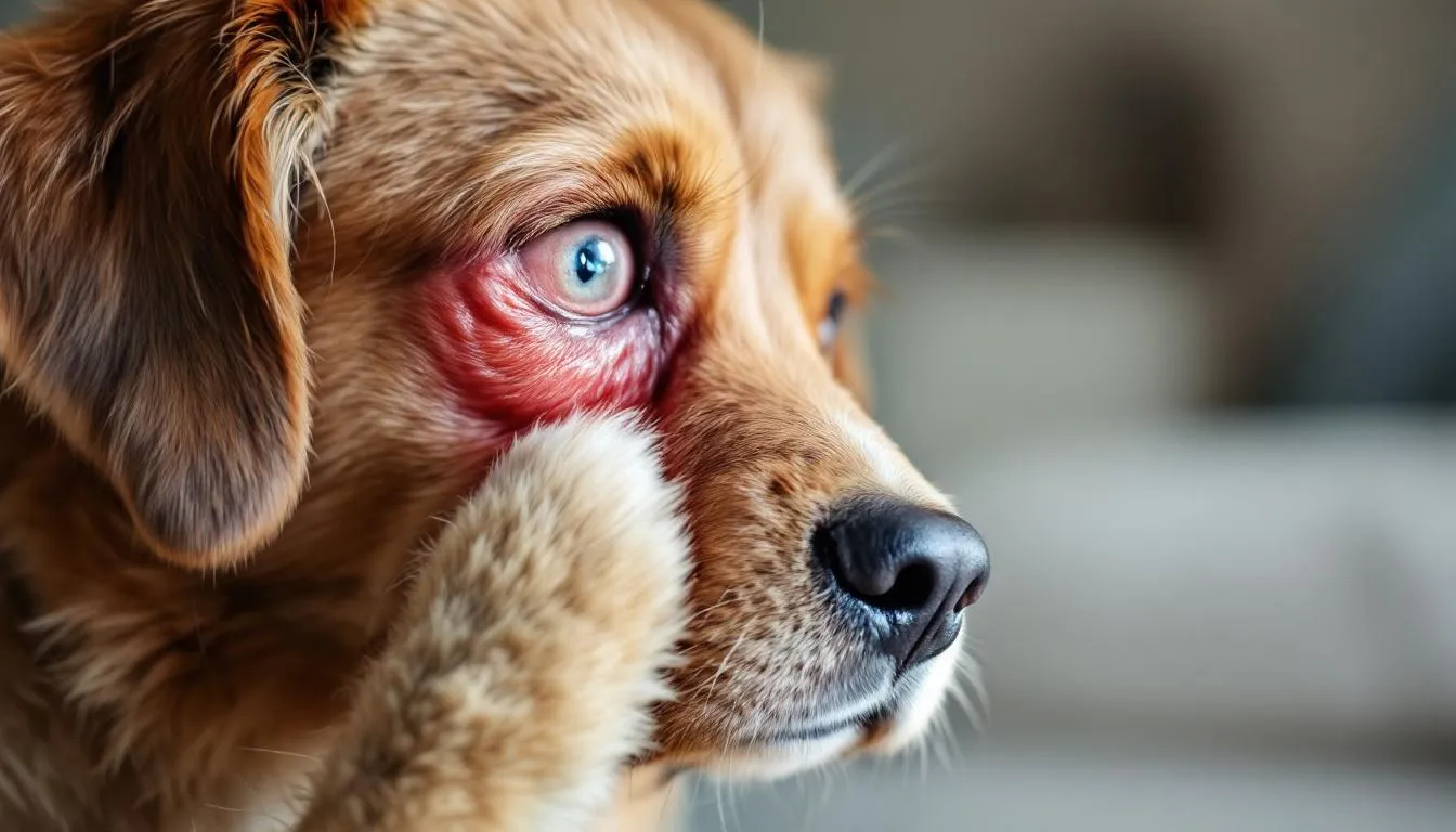
The clinical signs of conjunctivitis in dogs are typically quite distinctive and serve as important indicators for pet owners to recognize when their animals require veterinary attention. The most prominent symptom is conjunctival hyperemia, where the normally pink tissue becomes bright red due to increased blood flow and inflammation. Conjunctival hyperemia is a clinically observable manifestation of inflammation.
Ocular discharge represents another hallmark of the condition, with the character and color providing valuable diagnostic clues. Bacterial conjunctivitis typically produces thick, green or yellow discharge, while allergic or viral causes often result in clear or white secretions. The volume and consistency of this discharge can vary throughout the day and may cause the eyelids to stick together, particularly after periods of rest. Clinical lesions such as ulcerations or nodules may also be present and help guide diagnosis.
Dogs experiencing ocular discomfort from conjunctivitis frequently exhibit excessive blinking, squinting, or pawing at their affected eyes. This ocular pruritus drives many animals to rub their faces against furniture, carpeting, or other surfaces in an attempt to relieve the irritation. Such behavior can potentially worsen the condition by introducing additional contaminants or causing conjunctival trauma.
The presentation may be unilateral or bilateral conjunctivitis, depending on the underlying cause. While traumatic injuries or localized infections might affect only one eye initially, systemic conditions like allergies or viral infections typically result in bilateral involvement. Additional clinical signs may include conjunctival swelling, hair loss around the affected eyes, and in cases of concurrent systemic illness, accompanying respiratory symptoms such as nasal discharge or coughing. Subconjunctival hemorrhage, appearing as a bright red patch on the eye, can occur following trauma or systemic disease.
Types and Causes of Canine Conjunctivitis
Allergic Conjunctivitis
Allergic conjunctivitis affects dogs of all breeds but shows particular prevalence in animals predisposed to atopic dermatitis. This form of ocular disease results from exposure to environmental allergens including dust, pollen, molds, dust mites, perfumes, and grooming products. Food allergens and genetic predisposition to allergic responses also contribute significantly to the development of atopic conjunctivitis.
The condition typically manifests in young adult dogs, though any age can be affected. The classic triad of conjunctival hyperemia, conjunctival edema (chemosis), and ocular pruritus serves as the foundation for clinical diagnosis. Allergy testing is often performed to identify specific triggers. Conjunctival provocation tests can be used to confirm allergic responses and assess allergen sensitivity in clinical settings. Atopic dogs often develop this condition seasonally, corresponding to periods of increased environmental allergen exposure, particularly during spring and fall months when pollen counts are elevated.
Bacterial Conjunctivitis
Primary bacterial conjunctivitis occurs relatively rarely in dogs, with most cases representing secondary bacterial conjunctivitis that develops following compromise of the ocular surface. Secondary bacterial infection often exacerbates clinical lesions by intensifying tissue damage and inflammation. Common bacterial pathogens include Staphylococcus and Streptococcus species, which can proliferate when normal protective mechanisms are disrupted.
Several predisposing factors increase susceptibility to bacterial infection, including dry eye conditions that reduce tear production, corneal ulceration that compromises the ocular surface, eyelid abnormalities that alter normal blinking patterns, immune system disorders, and physical trauma. Certain breed-specific anatomical variations and parasitic infestations can also predispose dogs to developing secondary bacterial infections of the conjunctiva.
Viral Conjunctivitis
Viral conjunctivitis results from systemic viral infections that include ocular manifestations as part of their clinical presentation. The canine distemper virus represents one of the most significant viral causes, often producing severe conjunctivitis alongside other systemic symptoms. Canine herpesvirus-1 also commonly causes viral conjunctivitis, particularly in young dogs or those with compromised immune systems.
These viral forms of conjunctivitis can take considerably longer to resolve compared to bacterial cases, often requiring 3-4 weeks for complete healing. The contagious nature of these pathogens means that any dog breed can develop viral conjunctivitis upon exposure, making proper isolation and hygiene measures essential during treatment periods. Dogs diagnosed with viral conjunctivitis require isolation and supportive care to prevent spread and ensure recovery.
Ocular Diseases Associated with Conjunctivitis
Conjunctivitis in dogs often occurs alongside a variety of other ocular diseases, making a comprehensive clinical evaluation essential for accurate diagnosis and effective treatment. Conditions such as keratoconjunctivitis sicca (commonly known as dry eye), ulcerative keratitis, and uveitis can all present with similar clinical signs, including redness, ocular discharge, and ocular discomfort. Because these diseases can mimic or exacerbate conjunctivitis in dogs, a complete ophthalmic examination is necessary to distinguish between primary conjunctival inflammation and more complex ocular disease.
Bacterial conjunctivitis frequently develops as a secondary complication when the eye’s natural defenses are compromised by underlying issues like corneal ulcers or chronic dry eye. In these cases, addressing the primary ocular disease is crucial for resolving the conjunctivitis and preventing recurrence. Additionally, parasitic and fungal infections—such as those caused by Demodex mites or Thelazia eyeworms—can also lead to conjunctival inflammation. Diagnostic tools like conjunctival cytology and culture help veterinarians identify these less common causes and tailor treatment accordingly.
Systemic diseases can also manifest as conjunctivitis. For example, autoimmune disorders or neoplastic conditions may present with ocular lesions as part of a broader clinical picture. Dogs with atopic dermatitis are particularly prone to developing allergic conjunctivitis as a comorbidity, and managing the underlying allergy is key to controlling ocular symptoms. A thorough physical examination, along with bloodwork and biochemistry profiles, can help uncover these systemic diseases and guide a holistic treatment approach. By considering the full spectrum of potential causes, veterinarians can ensure that all contributing factors are addressed, leading to better outcomes for dogs affected by conjunctivitis and other ocular diseases.
Veterinary Diagnosis Process
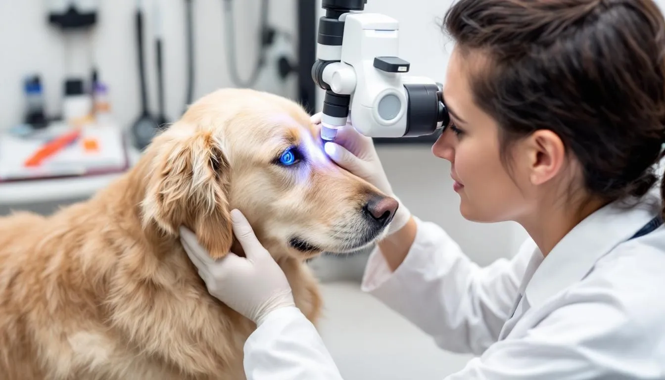
Accurate diagnosis of canine conjunctivitis requires a comprehensive approach that combines thorough clinical evaluation with appropriate diagnostic testing. The diagnostic process begins with a complete physical examination to assess the animal’s overall health status and identify any systemic diseases that might contribute to ocular symptoms. A nonspecific ocular lesion may be the initial presentation and requires thorough examination to rule out serious underlying disease.
A complete ocular examination forms the cornerstone of diagnosis, involving careful inspection of the eyelids, surrounding fur, eyelashes, third eyelid, and tear duct openings. The veterinarian will assess for eyelid abnormalities, foreign material, masses, or other ocular lesions that might contribute to the inflammatory process. Multiple bulbar conjunctival nodules may indicate parasitic infections such as onchocerciasis.
Schirmer tear test provides crucial information about tear production, as inadequate tear film can predispose to conjunctival inflammation and secondary bacterial infection. The Schirmer tear test involves placing a standardized strip of filter paper inside the lower eyelid for one minute to measure tear production; normal values are typically greater than 15 mm of wetting per minute. This helps identify cases of keratoconjunctivitis sicca (dry eye).
Fluorescein stain testing evaluates the integrity of the corneal surface, revealing corneal ulcers or other epithelial defects that might complicate the clinical picture. Additional diagnostic procedures may include measurement of intraocular pressure to rule out glaucoma, bacterial culture and sensitivity testing for persistent infections, conjunctival cytology to identify specific inflammatory patterns, and tissue biopsies in unusual cases. Conjunctival follicle formation observed during examination can indicate chronic or immune-mediated conjunctivitis. Other procedures may include allergy testing for recurrent allergic cases and tear duct flushing to assess drainage function.
This comprehensive diagnostic approach ensures that underlying conditions contributing to the conjunctivitis are identified and addressed, preventing recurrence and optimizing treatment outcomes.
Complications of Canine Conjunctivitis
When conjunctivitis in dogs is left untreated or inadequately managed, it can lead to a range of complications that significantly impact ocular health and overall quality of life. Chronic conjunctivitis is a common consequence, characterized by persistent redness, tearing, and ocular discomfort that can become increasingly difficult to resolve over time. This ongoing inflammation may also predispose the eye to more serious problems, such as corneal ulcers, especially in cases of frictional irritant conjunctivitis or following traumatic injury to the conjunctival tissue.
Corneal ulcers are particularly concerning, as they can cause severe pain and, if not promptly treated, may progress to deeper infections or even vision loss. In some cases, chronic or severe conjunctivitis can trigger additional complications like uveitis—an inflammation of the eye’s internal structures—or glaucoma, both of which can threaten vision and require urgent intervention. The risk of secondary bacterial or fungal infections also increases when the conjunctival barrier is compromised, further exacerbating clinical signs and complicating treatment.
To prevent these complications, regular veterinary check-ups and early intervention are essential. A complete ophthalmic examination and thorough clinical evaluation allow veterinarians to detect underlying issues, monitor for the development of corneal ulcers or other ocular diseases, and adjust treatment plans as needed. By addressing the root cause of conjunctivitis and providing timely, targeted therapy, pet owners and veterinarians can work together to alleviate ocular discomfort, prevent chronic conjunctivitis, and safeguard the long-term vision and well-being of affected dogs.
Treatment Options for Canine Conjunctivitis
Allergic Conjunctivitis Treatment
Management of allergic conjunctivitis focuses on controlling inflammation while identifying and minimizing exposure to triggering allergens. Topical corticosteroids represent the primary treatment modality for reducing conjunctival inflammation, though their use requires careful veterinary supervision to monitor for potential side effects.
Oral antihistamines provide systemic control of allergic responses and can be particularly beneficial for dogs with concurrent atopic dermatitis. When possible, allergen identification through allergy testing allows for more targeted environmental management strategies.
Supportive care measures include cold compresses to reduce swelling and discomfort, along with lubricating eye drops to maintain ocular surface moisture. Severe cases may require immunotherapy protocols designed to gradually desensitize the animal to specific allergens, providing long-term management for chronic allergic conjunctivitis.
Bacterial Conjunctivitis Treatment
Bacterial conjunctivitis responds well to appropriate antibiotic therapy, with topical applications generally preferred for localized infections. Commonly prescribed antibiotics include gentamicin, tobramycin, oxytetracycline, ciprofloxacin, and triple-antibiotic ointment formulations. The choice of specific antibiotic may be guided by bacterial culture and sensitivity testing, particularly in persistent or recurrent cases.
Severe infections may require oral antibiotic therapy in addition to topical treatment. Anti-inflammatory medications help reduce conjunctival swelling and associated ocular discomfort, improving patient comfort during the healing process. Treatment typically continues for 5-7 days, with most cases showing significant improvement within the first few days of appropriate therapy.
Viral Conjunctivitis Treatment
Viral conjunctivitis generally resolves through supportive care and time, as specific antiviral medications are rarely employed in veterinary practice. Treatment focuses on maintaining patient comfort and preventing secondary complications during the healing process.
Oral antioxidants may support immune system function, potentially accelerating recovery from viral infections. In select cases, antiviral medications might be considered, though their use remains limited in routine practice. Symptomatic treatment emphasizes maintaining ocular surface lubrication and managing any concurrent systemic illness that may accompany viral infections.
Specialized Treatments
Chronic dry eye conditions require specific management with immunomodulatory medications such as cyclosporine or tacrolimus, which stimulate natural tear production while reducing inflammation. These conditions typically require lifelong management with regular monitoring.
Anatomical eyelid abnormalities may necessitate surgical correction to restore normal eyelid function and prevent recurrent conjunctival irritation. Immune-mediated conjunctival diseases often require long-term immunosuppressive therapy, with treatment protocols tailored to individual patient needs and response patterns. Chronic inflammation can lead to epithelial conjunctival tissue proliferation, manifesting as irregular, raised lesions. Additionally, atypical pannus presents clinically with bilateral hyperemic thickening of the conjunctiva, multifocal follicle formation, and conjunctival depigmentation, requiring tailored immunosuppressive therapy.
Prevention and Management
Effective management of canine conjunctivitis extends beyond initial treatment to include measures that prevent recurrence and ensure complete healing. Elizabethan collars serve an important role during treatment by preventing rubbing and scratching that could worsen inflammation or introduce additional contaminants.
Regular veterinary rechecks allow monitoring of treatment response and adjustment of therapy as needed. Cases that fail to respond appropriately or show signs of worsening may require referral to veterinary ophthalmologists or dermatologists for specialized evaluation and management.
Early intervention represents a crucial factor in preventing the development of chronic conjunctivitis, which can be more challenging to manage and may result in permanent ocular changes. Vaccination programs help reduce the risk of viral conjunctivitis by protecting against common viral pathogens that can cause ocular disease.
Home Care and Remedies
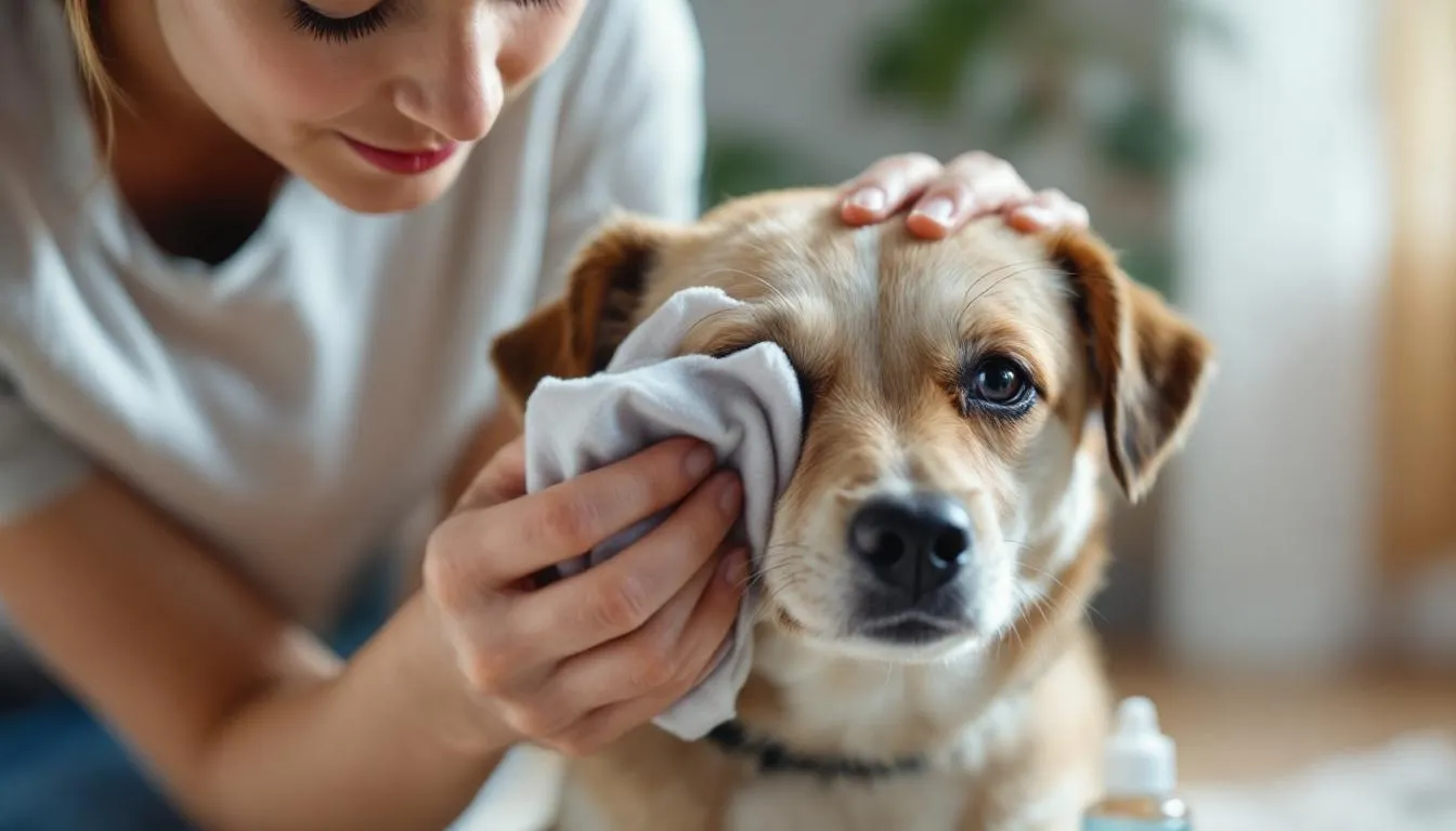
While professional veterinary care remains essential for proper diagnosis and treatment, certain home care measures can provide supportive benefits when used appropriately. Sterile saline eye washes can help remove discharge and flush minor irritants from the ocular surface, typically performed once or twice daily as directed by a veterinarian.
Eye flushing should only be performed after veterinary consultation to ensure that such measures will not cause additional injury or interfere with prescribed treatments. Some holistic products may help prevent tear duct blockage, though their efficacy varies and they should be discussed with a veterinarian before use.
It’s crucial to avoid using over-the-counter human eye medications without specific veterinary approval, as many formulations contain ingredients that can be toxic to dogs or may worsen certain types of conjunctivitis. Always discuss any proposed home treatments with your veterinarian to ensure they complement rather than interfere with prescribed therapy.
Recovery Timeline and Prognosis
The recovery timeline for canine conjunctivitis varies significantly depending on the underlying cause and severity of the condition. Bacterial conjunctivitis typically shows rapid improvement with appropriate antibiotic treatment, with most cases resolving within 5-7 days of initiating therapy.
Viral conjunctivitis requires considerably more patience, often taking 3-4 weeks for complete recovery. During this extended period, supportive care and monitoring for secondary complications remain important components of management.
Allergic conjunctivitis presents ongoing challenges, as symptoms may persist until triggering allergens are identified and controlled. Seasonal allergies may show cyclical patterns, requiring anticipatory treatment during high-risk periods.
Chronic conditions such as dry eye require lifelong management, with prognosis depending on the underlying cause and response to treatment. Most dogs with properly managed dry eye can maintain comfortable vision and good quality of life with appropriate ongoing care.
The overall prognosis for canine conjunctivitis remains favorable when appropriate treatment is instituted promptly. Severe or chronic cases may have more guarded outcomes, particularly when underlying systemic or intraocular disease complicates the clinical picture.
Contagiousness and Prevention
The contagious nature of conjunctivitis varies significantly depending on the underlying cause. Viral and bacterial forms often demonstrate high contagiousness between dogs, requiring isolation measures during active infection periods to prevent spread to other animals.
Viral conjunctivitis, while highly contagious among dogs, does not typically pose transmission risks to humans. However, bacterial forms can potentially spread to humans through direct contact, making proper hygiene essential when handling affected animals.
Allergic conjunctivitis, immune-mediated forms, and dry eye conditions are not contagious and do not require isolation measures. These conditions result from individual susceptibility factors rather than infectious agents.
Proper hygiene practices, including thorough hand washing after handling affected animals and avoiding sharing of bedding or toys between infected and healthy dogs, help minimize disease transmission. Vaccination against common viral pathogens provides additional protection against viral causes of conjunctivitis.
When to Seek Emergency Care
Certain presentations of conjunctivitis warrant immediate veterinary attention due to the risk of severe ocular diseases or complications. Sudden onset of severe eye pain or vision changes may indicate more serious intraocular disease requiring emergency intervention.
Thick, purulent discharge accompanied by systemic signs of illness such as fever, lethargy, or respiratory distress suggests possible systemic infection that may require aggressive treatment. The development of corneal ulceration or corneal cloudiness indicates compromise of the corneal surface that could threaten vision if not addressed promptly.
Signs suggestive of increased intraocular pressure, such as a enlarged or firm eye, severe pain, or sudden vision loss, may indicate secondary glaucoma requiring immediate treatment to preserve vision. Failure to respond to prescribed treatment within the expected timeframe or development of worsening symptoms during treatment also warrants emergency evaluation.
Any development of secondary complications, such as extensive corneal involvement or signs of deeper ocular infection, requires prompt professional assessment to prevent permanent damage to vision or ocular structures.
FAQ
Can conjunctivitis in dogs resolve without treatment?
While some very mild cases of conjunctivitis might improve naturally in dogs with robust immune systems, most cases require veterinary treatment to prevent complications and ensure complete recovery. Untreated conjunctivitis carries significant risks including chronic pain, corneal scarring, persistent infections, and potential vision problems. The inflammatory process can worsen over time without appropriate intervention, making early treatment essential for optimal outcomes. Even cases that appear to improve without treatment may develop into chronic conditions that are more difficult to manage long-term.
How can I tell if my dog’s conjunctivitis is bacterial or viral?
Distinguishing between bacterial and viral conjunctivitis requires careful observation of clinical signs and often veterinary testing. Bacterial conjunctivitis typically produces thick, green or yellow discharge and usually responds quickly to antibiotic treatment within 5-7 days. Viral conjunctivitis often presents with clearer discharge and takes considerably longer to resolve, typically 3-4 weeks, and may be accompanied by other respiratory symptoms like nasal discharge or coughing. However, definitive diagnosis requires veterinary examination and potentially laboratory testing including conjunctival cytology or bacterial cultures for accurate identification.
Is it safe to use human eye drops on my dog?
Never use human eye medications on dogs without explicit veterinary approval, as many ingredients that are safe for humans can be toxic to dogs or may worsen existing eye conditions. Some human eye drops contain preservatives, vasoconstrictors, or other compounds that can cause severe reactions in dogs. Sterile saline solution is generally safe for cleaning purposes but should not replace proper veterinary treatment. Always consult your veterinarian before using any eye products on your pet, as they can recommend appropriate veterinary-specific formulations that are both safe and effective for your dog’s particular condition.
Why does my dog keep getting conjunctivitis?
Recurring conjunctivitis often indicates an underlying condition that predisposes your dog to repeated episodes. Common causes include allergies (particularly atopic dermatitis), chronic dry eye, anatomical abnormalities like eyelid malformations, immune system disorders, or chronic bacterial infections. Environmental factors such as exposure to irritants or allergens may also contribute to repeated episodes. A comprehensive diagnostic workup including allergy testing, tear production measurement, and evaluation for anatomical abnormalities may be necessary to identify the root cause. Once identified, long-term management strategies can often prevent future occurrences and improve your dog’s comfort.
How long should I keep my dog isolated if they have infectious conjunctivitis?
Isolation periods depend on the specific type of infectious conjunctivitis diagnosed. Dogs with bacterial conjunctivitis should typically be isolated until 24-48 hours after starting appropriate antibiotic treatment, when they are no longer considered contagious. Viral conjunctivitis may require longer isolation periods of 1-2 weeks or until symptoms completely resolve, as viral shedding can continue throughout the illness. Your veterinarian will provide specific recommendations based on the diagnosed cause and your dog’s response to treatment. Practice good hygiene including thorough hand washing after handling affected dogs to prevent spread to other pets or potential transmission of bacterial forms to humans.


