


Key Takeaways
- Canine cataracts are lens opacities that cause vision impairment and can lead to complete blindness if left untreated
- Hereditary factors and diabetes mellitus are the two most common causes of cataracts in dogs, with 75-80% of diabetic dogs developing cataracts within one year
- Cataract surgery (phacoemulsification), also known as cataract removal, is the only effective treatment to restore vision, with success rates of 80-90% when performed early
- Cataract removal (surgical removal of the lens) is the primary way cataracts are treated in dogs, typically performed by a veterinary ophthalmologist. Medications may be used to manage inflammation and prevent complications, but surgery is the most effective option for restoring vision.
- Early intervention is crucial as mature and hypermature cataracts increase surgical complications and reduce success rates
- Untreated cataracts can cause painful secondary conditions including lens-induced uveitis, glaucoma, and retinal detachment
Canine cataracts are lens opacities that cause vision impairment and can lead to complete blindness if left untreated
Hereditary factors and diabetes mellitus are the two most common causes of cataracts in dogs, with 75-80% of diabetic dogs developing cataracts within one year
Cataract surgery (phacoemulsification), also known as cataract removal, is the only effective treatment to restore vision, with success rates of 80-90% when performed early
Cataract removal (surgical removal of the lens) is the primary way cataracts are treated in dogs, typically performed by a veterinary ophthalmologist. Medications may be used to manage inflammation and prevent complications, but surgery is the most effective option for restoring vision.
Early intervention is crucial as mature and hypermature cataracts increase surgical complications and reduce success rates
Untreated cataracts can cause painful secondary conditions including lens-induced uveitis, glaucoma, and retinal detachment

What Are Canine Cataracts?
When your dog’s once-bright eyes develop a cloudy, white appearance, you’re likely witnessing the formation of cataracts—a condition that affects thousands of dogs annually and can progress to complete blindness without proper intervention. Dog cataracts represent an opacity or clouding of the eye's lens, the transparent structure responsible for focusing light onto the retina to create clear vision.
In a healthy dog’s eye, the eye's lens focuses light precisely onto the retina, much like a camera lens focuses light onto film. The lens achieves this through its crystal-clear transparency and precise protein arrangement. The eye's lens helps focus light onto the retina, enabling clear vision. When cataracts form, this transparency becomes compromised as lens proteins clump together or water balance changes, causing light to scatter rather than focus properly.
It’s crucial to distinguish cataracts from lenticular sclerosis, also known as nuclear sclerosis, a normal aging change that causes a bluish-gray haze in older dogs’ eyes. While lenticular sclerosis may look concerning to owners, it typically doesn’t significantly impact the dog’s vision. Only a veterinary examination can reliably differentiate between these conditions, as the distinction affects treatment decisions and prognosis.
The impact on your dog’s quality of life depends on the cataract’s severity and progression rate. Dogs with cataracts experience diminished vision as the lens becomes more opaque. Early-stage cataracts may cause minimal vision impairment, while a complete cataract can render dogs completely blind, affecting their ability to navigate familiar environments, play, and maintain their normal activity levels.
As dogs cataracts progress, the lens begins to change, and vision impairment increases.
Causes of Cataracts in Dogs
Understanding why cataracts develop helps owners recognize risk factors and take preventive measures when possible. The causes of dogs cataracts vary significantly, with some being entirely preventable while others stem from genetic predisposition. A significant percentage of dogs affected by cataracts develop the condition due to hereditary factors or underlying diseases such as diabetes mellitus.
Hereditary cataracts represent the most common cause in domesticated dogs, affecting over 20 dog breeds with varying degrees of severity and age of onset. American Cocker Spaniels, Labrador Retrievers, Bichon Frise, Boston Terriers, and Siberian Huskies show particularly high predisposition rates. These inherited cataracts can appear in young dogs or develop gradually as the dog ages, depending on the specific genetic mutation involved.
Diabetes mellitus (also known as sugar diabetes) creates a particularly urgent scenario, as diabetic dogs develop cataracts with alarming frequency and speed. The mechanism involves excess glucose in the eye converting to sorbitol, which draws water into the lens and causes rapid swelling and opacity formation. This process can occur literally overnight in some cases, transforming a seeing dog into a blind one within days.
Traumatic cataracts result from blunt force injuries, penetrating wounds, or lens laceration during eye trauma. Even seemingly minor injuries can disrupt the lens capsule, initiating cataract formation that may progress slowly over months or years.
Secondary cataracts develop as complications of other eye diseases, including chronic uveitis, glaucoma, and progressive retinal atrophy. The deep inflammation associated with these conditions can damage lens fibers and accelerate cataract formation.
Less common causes include nutritional deficiencies in puppies fed improper milk replacement formulas, radiation exposure during cancer treatments, and congenital cataracts present at birth due to developmental abnormalities during fetal growth.
Stages and Classification of Cataracts
Veterinary ophthalmologists classify cataracts based on their extent and maturity, which directly impacts treatment decisions and surgical outcomes. Understanding these stages helps owners appreciate the importance of early intervention.
Incipient cataracts affect less than 15% of the lens area and typically don’t significantly impair vision. Dogs with incipient cataracts may show no behavioral changes, and the condition might only be detected during routine veterinary examinations. These early-stage cataracts require monitoring but don’t necessarily need immediate surgical intervention.
Immature cataracts involve 15-99% of the lens, creating variable vision impairment depending on their location and density. Dogs may begin showing difficulty with depth perception, hesitation on stairs, or reduced confidence in low-light situations. The lens focuses light unevenly, creating a partially obscured visual field.
Mature cataracts represent complete lens opacity where the cataract completely blocks light transmission to the retina. Dogs with mature cataracts experience near-complete blindness, though they may still detect bright light and shadows. At this stage, the entire lens appears white or gray when examined.
Hypermature cataracts develop when lens proteins begin breaking down, causing the lens to shrink and potentially wrinkle. As the lens begins to shrink and wrinkle, the lens capsule appears wrinkled, and these advanced cataracts carry increased risks of complications including severe lens-induced uveitis and secondary glaucoma. Chronic cataracts, if left untreated, can lead to secondary complications such as glaucoma and retinal detachment, making timely surgery important to prevent these issues.
Cataracts are also classified by location within the lens. Nuclear cataracts affect the lens center, cortical cataracts involve the outer lens layers, and subcapsular cataracts form near the lens capsule. The location influences how quickly vision deteriorates and affects surgical complexity.
Signs and Symptoms of Cataracts
Recognizing the early signs of cataracts enables prompt veterinary intervention, potentially preserving more vision and reducing surgical complications. The symptoms vary depending on the cataract’s stage and progression rate.
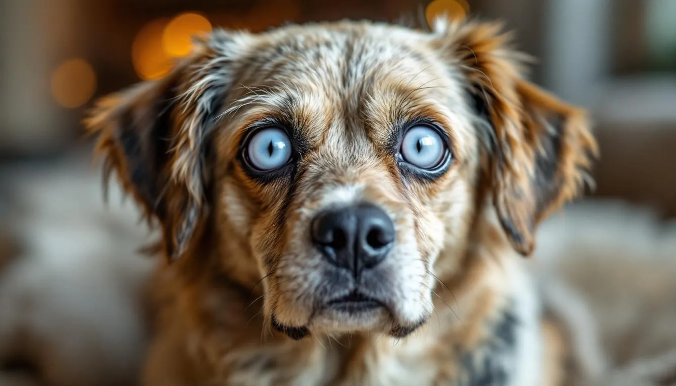
The most obvious sign is progressive cloudiness or white appearance in the pupil area. Rather than the normal black pupil, owners notice a white, gray, or bluish discoloration that becomes more pronounced over time. This change is often most visible in certain lighting conditions or when photographing the dog with flash.
Behavioral changes often provide the earliest clues before visible opacity appears. Dogs may begin bumping into furniture, hesitating at the top or bottom of stairs, or showing reluctance to navigate in dim lighting. Many dogs develop compensatory behaviors, staying closer to walls or moving more cautiously through familiar spaces.
Activity level changes frequently accompany vision loss. Previously active dogs may become less interested in playing fetch, show reluctance to jump onto furniture, or display anxiety in new environments. Some dogs become more clingy, staying closer to their owners for guidance and reassurance.
Secondary signs can indicate complications requiring urgent attention. Eye redness, discharge, squinting, or signs of pain suggest the development of lens-induced uveitis or secondary glaucoma. These complications can threaten not only vision but the comfort and health of the entire eye.
When only one eye is affected initially, dogs often compensate remarkably well, and owners may not notice vision problems until the condition progresses significantly or affects both eyes. This underscores the importance of regular veterinary examinations for early detection.
Diagnosis of Canine Cataracts
Accurate diagnosis requires professional veterinary examination, as several conditions can mimic cataracts or occur simultaneously. Veterinarians use specialized examinations and tests to diagnose cataracts in dogs, ensuring accurate identification and differentiation from other eye conditions. The diagnostic process involves multiple steps to determine not only the presence of cataracts but also their impact on vision and overall eye health.
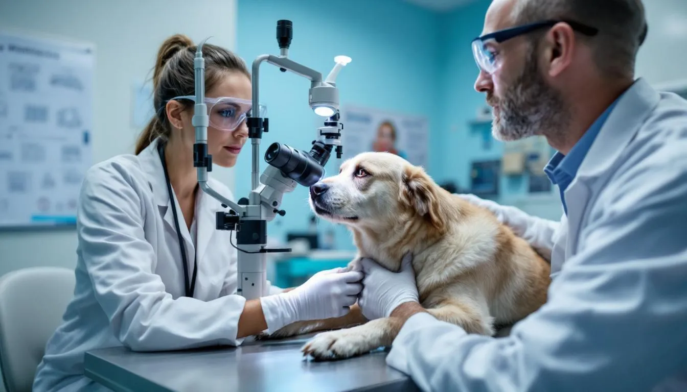
A comprehensive ophthalmic examination begins with external inspection of the eyes, eyelids, and surrounding tissues. The veterinarian or veterinary ophthalmologist uses specialized instruments including an ophthalmoscope and slit lamp biomicroscopy to examine internal eye structures in detail.
Pupil dilation allows complete visualization of the lens and retina. Special eye drops temporarily enlarge the pupils, enabling examination of the entire lens surface and detection of early cataracts that might not be visible through an undilated pupil. This process is painless but requires keeping the dog calm during examination.
Tonometry measures intraocular pressure to detect glaucoma, which can either cause or result from cataracts. Elevated eye pressure requires immediate treatment to prevent permanent vision loss and pain.
Additional diagnostic tests may include Schirmer tear tests to evaluate tear production, fluorescein staining to check for corneal damage, and ocular ultrasound when cataracts prevent clear visualization of internal eye structures.
For surgical candidates, advanced diagnostics become essential. Electroretinogram (ERG) testing evaluates retinal function to ensure the retina can respond to light once cataracts are removed. Dogs with poor retinal function may not benefit from surgery, making this test crucial for determining surgical candidacy.
The examination also focuses on differentiating cataracts from nuclear sclerosis and identifying any concurrent eye diseases that might affect treatment decisions or surgical outcomes.
Dog Breeds and Cataracts
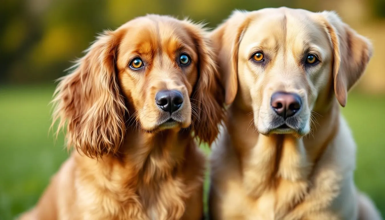
Cataracts in dogs can affect any breed, but certain dog breeds are especially prone to developing cataracts due to hereditary factors. Hereditary cataracts are a significant concern in veterinary ophthalmology, as they can appear in many dog breeds and at various stages of a dog’s life. Breeds such as the American Cocker Spaniel, Labrador Retriever, French Poodle, Boston Terrier, and Welsh Springer Spaniel are particularly susceptible to cataract formation, often due to inherited genetic mutations that affect the lens.
In these breeds, hereditary cataracts may develop early in life or as the dog ages, and they can affect one or both eyes. Some breeds are also more likely to experience specific types of cataracts. For example, the Bichon Frise and Boston Terrier are known to develop nuclear sclerosis, an age-related change that causes a bluish haze in the lens, while breeds like the Labrador Retriever and German Shepherd are at higher risk for lens luxation, where the lens becomes dislocated. These breed-specific tendencies highlight the importance of understanding your dog’s genetic background and monitoring for early signs of vision loss.
Diabetic cataracts are another major concern, especially in breeds already predisposed to diabetes mellitus. Approximately 75-80% of diabetic dogs will develop cataracts within the first year of diagnosis, regardless of how well their diabetes is managed. This rapid cataract progression can lead to sudden vision loss and increases the risk of complications such as severe lens induced uveitis if left untreated.
Regular eye exams by a veterinary ophthalmologist are essential for dogs in high-risk breeds, as early detection of cataracts can make a significant difference in treatment outcomes. Early intervention, including timely cataract surgery, can help restore a dog’s vision and prevent secondary complications like untreated cataracts progressing to glaucoma or retinal detachment. Veterinary ophthalmologists can also help distinguish between hereditary cataracts, nuclear sclerosis, and other lens disorders, ensuring that your dog receives the most appropriate care.
While breed plays a significant role in cataract risk, other factors such as age, overall health, and diet also contribute to cataract development. As dogs age, the likelihood of cataract formation increases, and conditions like diabetes mellitus further elevate this risk. Maintaining a healthy lifestyle, providing balanced nutrition, and scheduling regular veterinary check-ups can help reduce the risk of cataracts and support your dog’s overall eye health.
For breeders, understanding the hereditary nature of cataracts in many dog breeds is crucial. Responsible breeding practices, including genetic screening and avoiding breeding dogs diagnosed with hereditary cataracts, can help reduce the incidence of cataracts in future generations.
Ultimately, while some breeds are more prone to cataracts, any dog can develop cataracts at any age. By staying vigilant for changes in your dog’s eyes, seeking prompt veterinary attention, and working closely with veterinary ophthalmologists, you can help protect your dog’s vision and quality of life. Early detection and treatment, including cataract surgery when appropriate, offer the best chance for maintaining your dog’s sight and preventing complications like severe lens induced uveitis or permanent vision loss.
Treatment Options for Canine Cataracts
Treatment approaches for canine cataracts depend on the stage of development, underlying causes, and the dog’s overall health status. While surgical removal remains the only definitive cure, various management strategies can help maintain comfort and slow progression in certain cases.
Monitoring and observation suits dogs with early incipient cataracts that don’t significantly impact vision. Regular examinations track progression and help determine optimal timing for intervention if cataracts advance.
Medical management focuses on controlling inflammation and preventing complications rather than reversing cataracts. Anti-inflammatory eye drops help manage secondary inflammation and may reduce the risk of developing glaucoma. These medications don’t cure cataracts but can maintain eye comfort and health. Topical therapy, such as anti-inflammatory eye drops, can help manage symptoms but does not reverse cataracts. Topical aldose reductase inhibitors have been studied for their potential to delay cataract progression, especially in diabetic dogs, but are not a reliable treatment.
For diabetic dogs, some veterinarians recommend antioxidant supplements like those containing vitamin C and E, though evidence for preventing cataract progression remains limited. These supplements work best when started early in the diabetic process, before significant cataracts develop.
Cataract dissolution, or the natural breakdown of cataracts, is rare and often leads to complications such as deep inflammation.
Surgical intervention represents the only treatment capable of restoring vision. Cataract surgery involves removing the clouded lens and typically replacing it with an artificial lens to restore focusing ability. The decision for surgery depends on multiple factors including the dog’s age, overall health, stage of cataracts, and owner commitment to post-operative care.
Management of underlying conditions is crucial for dogs with diabetes mellitus or other systemic diseases contributing to cataract formation. While controlling diabetes doesn’t reverse existing cataracts, it may slow progression and improve overall health for surgical candidates.
For dogs who aren’t surgical candidates or whose owners choose not to pursue surgery, environmental modifications can significantly improve quality of life. These adaptations help blind or vision-impaired dogs navigate safely and maintain independence.
Cataract Surgery in Dogs
Canine cataract surgery, also known as cataract removal, represents one of the most successful procedures in veterinary ophthalmology, with success rates of 80-90% for restoring functional vision when performed by experienced veterinary ophthalmologists. The procedure, called phacoemulsification, uses sophisticated techniques adapted from human cataract surgery.
The surgical process begins with general anesthesia and careful positioning of the dog’s head. The surgeon creates a small incision in the cornea and uses high-frequency ultrasonic vibrations to break up the cataractous lens material. This emulsified lens material is then aspirated from the eye, leaving the lens capsule intact.
Artificial lens implantation typically follows lens removal to restore the eye’s focusing ability. Without an artificial lens, dogs can see but lack sharp focus for detailed vision. Modern intraocular lenses designed specifically for dogs provide excellent visual outcomes and long-term stability.
Pre-surgical evaluation is extensive and crucial for success. Dogs undergo complete physical examinations, blood work to detect systemic diseases, and specialized eye tests including ERG to confirm retinal function. Control of any existing eye inflammation is essential before surgery, often requiring weeks of medical treatment.
Same-day bilateral surgery is often recommended when both eyes have cataracts, as it reduces anesthesia exposure and allows for coordinated recovery. However, some cases may require staged procedures depending on the dog’s health and specific circumstances.
The timing of surgery significantly impacts outcomes. Early intervention, before cataracts become mature or hypermature, reduces complication rates and improves visual results. Once cataracts progress to advanced stages, surgical risks increase and success rates may decline. Successful cataract removal can significantly restore a dog's sight, greatly improving quality of life.
Surgery Costs and Considerations
Understanding the financial commitment helps owners plan for cataract surgery and explore available options. The total cost varies based on geographic location, complexity of the case, and presence of complications.
Initial consultation with a veterinary ophthalmologist typically costs $200-$300 and includes comprehensive eye examination and preliminary surgical assessment. This appointment determines surgical candidacy and provides cost estimates specific to your dog’s situation.
Pre-surgical diagnostics including ERG testing, ocular ultrasound, blood work, and specialized imaging add $1,000-$1,200 to total costs. These tests are essential for identifying dogs who won’t benefit from surgery and detecting conditions that might complicate the procedure.
Bilateral cataract surgery ranges from $2,700-$4,000 depending on the surgical facility and case complexity. This includes the procedure itself, intraocular lens implants, initial post-operative medications, and immediate follow-up care.
Total average costs around $3,500 represent a significant investment, but many owners find the restoration of their dog’s sight invaluable for quality of life. Some veterinary schools and specialty practices offer payment plans or reduced-cost options for qualifying cases.
Pet insurance coverage varies significantly between policies and companies. Hereditary conditions may be excluded unless coverage was in place before symptom onset, while traumatic cataracts are more likely to be covered.
Post-Surgical Care and Recovery
Successful cataract surgery recovery requires dedicated owner commitment and strict adherence to post-operative protocols. The healing process typically takes several weeks to months, with most dogs showing dramatic vision improvement within days of surgery.
Immediate post-operative period involves overnight hospitalization for monitoring and pain management. Dogs receive medications to control inflammation, prevent infection, and manage discomfort. Most dogs are alert and comfortable within hours of surgery completion.
Home care requirements include administering prescribed eye drops 2-4 times daily for several weeks. These medications prevent infection, control inflammation, and promote healing. Consistent medication timing and proper administration technique are crucial for preventing complications.
Activity restriction is essential during the initial healing period. Dogs must remain on leash for all outdoor activities, with no running, jumping, or rough play allowed. Swimming and activities that might result in eye trauma are prohibited until healing is complete.
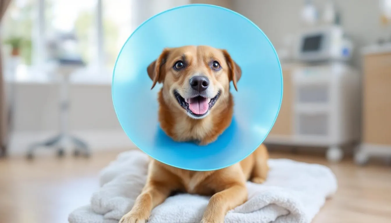
Protective measures include wearing an Elizabethan collar or inflatable cone to prevent scratching or rubbing the surgical sites. While dogs often dislike these devices, they’re essential for preventing self-trauma that could compromise surgical results.
Follow-up appointments occur frequently during the first few weeks, then gradually become less frequent as healing progresses. These visits allow the veterinary ophthalmologist to monitor healing, adjust medications, and detect early signs of complications.
Recovery timeline varies between individuals, but most dogs show significant vision improvement within 3-7 days. Complete healing may take 6-8 weeks, during which activity restrictions gradually lift based on healing progress.
Signs requiring immediate veterinary attention include increased redness, discharge, squinting, or behavioral changes suggesting pain or vision loss. Early intervention for complications often prevents serious long-term problems.
Complications of Untreated Cataracts
Allowing cataracts to progress without treatment creates risks extending far beyond simple vision loss. Advanced cataracts can lead to painful complications that threaten not only sight but the comfort and health of the entire eye.
Lens-induced uveitis represents one of the most serious complications of mature and hypermature cataracts. As lens proteins leak into the eye, they trigger severe inflammation that causes pain, redness, and further vision impairment. Without treatment, this inflammation can become chronic and extremely difficult to control.
Secondary glaucoma develops when inflammation or lens swelling increases intraocular pressure beyond safe levels. Elevated eye pressure causes pain and can permanently damage the optic nerve, leading to irreversible blindness even if cataracts are later removed.
Lens luxation occurs when the lens dislocates from its normal position within the eye. Hypermature cataracts weaken the structures holding the lens in place, potentially causing the lens to move forward into the anterior chamber or backward into the vitreous cavity.
Retinal detachment can result from chronic inflammation or sudden changes in eye pressure. Once the retina detaches, surgical repair becomes complex and expensive, with variable success rates for restoring vision.
Endophthalmitis represents the most severe complication, involving infection within the eye that can lead to loss of the entire eye. This condition requires aggressive treatment and may necessitate surgical removal of the eye to prevent spread of infection.
Chronic pain accompanies many of these complications, significantly impacting quality of life. Dogs may show signs of discomfort including pawing at the eye, head shaking, or behavioral changes suggesting chronic pain.
The progression to hypermature cataracts also increases surgical complexity and reduces success rates if surgery is later attempted. Prevention through early intervention remains far superior to treating advanced complications.
Special Considerations for Diabetic Dogs
Diabetic dogs face unique challenges regarding cataract development and management. The relationship between diabetes mellitus and cataracts is so strong that cataract formation often serves as the first sign alerting owners to their dog’s diabetic condition.
Rapid progression characterizes diabetic cataracts, with 75-80% of diabetic dogs developing cataracts within the first year of diagnosis. Unlike hereditary cataracts that may develop slowly over years, diabetic cataracts can form within days or weeks, creating urgent situations requiring immediate veterinary attention.
Formation mechanism involves excess glucose in the blood and eye fluids converting to sorbitol through enzymatic processes. Sorbitol accumulates within lens fibers and draws water into the lens, causing swelling and protein disruption that creates opacity.
Intumescent cataracts develop when rapid water influx causes the lens to swell significantly, creating characteristic “waterclefts” visible on examination. These swollen cataracts carry high risks of lens capsule rupture and severe lens-induced uveitis.
Higher complication rates occur in diabetic dogs, particularly regarding severe lens-induced uveitis and secondary glaucoma. The inflammatory response to diabetic cataracts tends to be more intense and harder to control than cataracts from other causes.
Surgical urgency often necessitates earlier intervention in diabetic dogs to prevent irreversible complications. Waiting for cataracts to “mature” may not be appropriate when rapid progression and high inflammation risk are present.
Continued progression occurs regardless of how well diabetes is controlled once cataracts begin forming. While excellent diabetic management may slow progression slightly, it cannot prevent or reverse existing cataracts.
Emergency situations can develop rapidly in diabetic dogs, particularly when lens capsule rupture releases inflammatory proteins into the eye. These cases may require immediate surgery or intensive medical management to preserve the eye and prevent permanent damage.
Living with Cataracts: Management and Quality of Life
When surgery isn’t possible or owners choose conservative management, numerous strategies can help dogs maintain good quality of life despite vision impairment. Dogs adapt remarkably well to gradual vision loss, often surprising owners with their resilience and adaptability.
Environmental modifications form the foundation of supporting vision-impaired dogs. Maintaining consistent furniture arrangements prevents confusion and reduces collision risks. Clear pathways through the house and removing obstacles like toys or decorative items from walking areas enhance navigation safety.
Verbal cues and consistent routines become essential communication tools. Teaching commands like “step up,” “step down,” and “careful” helps guide dogs through challenging terrain. Consistent daily routines for feeding, walks, and bathroom breaks provide security and predictability.
Safe exercise areas ensure continued physical activity without injury risks. Fenced yards or long leashes in familiar areas allow dogs to explore while preventing dangerous situations. Swimming can provide excellent exercise for dogs comfortable in water.
Monitoring for behavioral changes helps identify dogs struggling with vision loss. Some dogs develop anxiety, depression, or fear-based behaviors requiring professional behavior modification or anti-anxiety medications.
Regular veterinary monitoring remains crucial even without surgical intervention. Routine examinations detect developing complications requiring treatment, such as glaucoma or uveitis that could cause pain even if vision can’t be restored.
Quality of life assessment should be ongoing, considering factors like appetite, activity level, social interaction, and apparent comfort. Most dogs with gradual vision loss maintain excellent quality of life with appropriate support and environmental modifications.
The decision between surgical intervention and medical management depends on numerous factors including the dog’s age, health status, owner resources, and individual circumstances. Neither choice is inherently superior—the best approach varies for each dog and family situation.
Prevention and Breeding Considerations
While not all cataracts are preventable, understanding risk factors and implementing screening programs can significantly reduce the incidence of hereditary cataracts in future generations.
Genetic screening programs for breeding dogs help identify carriers of cataract-causing mutations. Many dog breeds now have available genetic tests that can identify dogs carrying genes for hereditary cataracts, even if the dogs themselves haven’t developed cataracts.
Eye certification programs conducted by veterinary ophthalmologists provide annual examinations for breeding dogs. Organizations like the Orthopedic Foundation for Animals maintain databases of eye examination results, helping breeders make informed decisions about which dogs to include in breeding programs.
Responsible breeding practices involve avoiding breeding dogs diagnosed with hereditary cataracts or dogs that have produced offspring with early-onset cataracts. This requires long-term commitment from breeders to track the health of puppies throughout their lives.
Early diabetes detection and management can delay cataract formation in susceptible dogs. Regular veterinary examinations including blood glucose monitoring help identify diabetes before severe symptoms develop.
Proper nutrition during development is crucial for puppies, particularly regarding appropriate milk replacement formulas if hand-feeding becomes necessary. Nutritional deficiencies during critical growth periods can predispose to cataract formation.
Eye protection during radiation therapy or other medical treatments that might increase cataract risk should be discussed with veterinary oncologists when planning cancer treatments.
Regular eye examinations for at-risk breeds enable early detection and intervention. Annual eye exams by veterinary ophthalmologists can identify developing cataracts before they significantly impact vision.
FAQ
Can cataracts in dogs be prevented with eye drops or supplements?
No topical eye drops can prevent or reverse cataracts. While some antioxidant supplements may help delay diabetic cataracts, surgery remains the only definitive treatment to restore vision. Eye drops can help manage secondary inflammation and prevent glaucoma, but they don’t affect the cataract itself.
How quickly do cataracts progress in dogs?
Progression varies significantly by cause and individual. Diabetic cataracts can develop rapidly within days to weeks, while hereditary cataracts may progress slowly over months to years. Some dogs maintain partial vision for extended periods, while others may lose vision quickly once cataracts begin forming.
Is my senior dog too old for cataract surgery?
Age alone doesn’t disqualify dogs from surgery. The decision depends on overall health, anesthetic risk, and ability to comply with post-operative care. Many senior dogs successfully undergo surgery with excellent outcomes. A thorough pre-surgical evaluation helps determine if your dog is a good candidate regardless of age.
What’s the difference between cataracts and the blue haze I see in my older dog’s eyes?
The blue haze is likely nuclear sclerosis, a normal aging change that rarely affects vision significantly. Nuclear sclerosis occurs in most dogs over 7 years old and causes lens hardening without opacity. Cataracts appear as white or gray opacities and progressively impair vision. Only a veterinarian can definitively distinguish between the two conditions.
Can dogs live happily without vision if surgery isn’t possible?
Yes, dogs adapt remarkably well to blindness, especially when it develops gradually. With environmental modifications, consistent routines, and proper care, blind dogs can maintain excellent quality of life. They rely more heavily on smell and hearing to navigate their world, often surprising owners with their adaptability and continued enjoyment of activities like walks and play.


